|
|
|
|
|
Fig.1 |
Fig.2 |
Fig.3 |
 |
 |
 |
| Fig.4 Fig.5 Fig.6 | Fig.7 Fig.8 Fig.9 | Fig.10 Fig.11 Fig.12 |
|
|
|
|
|
Fig.1 |
Fig.2 |
Fig.3 |
|
|
|
| Fig.4 Fig.5 Fig.6 | Fig.7 Fig.8 Fig.9 | Fig.10 Fig.11 Fig.12 |
Dental Education Lecture: Central Cusp
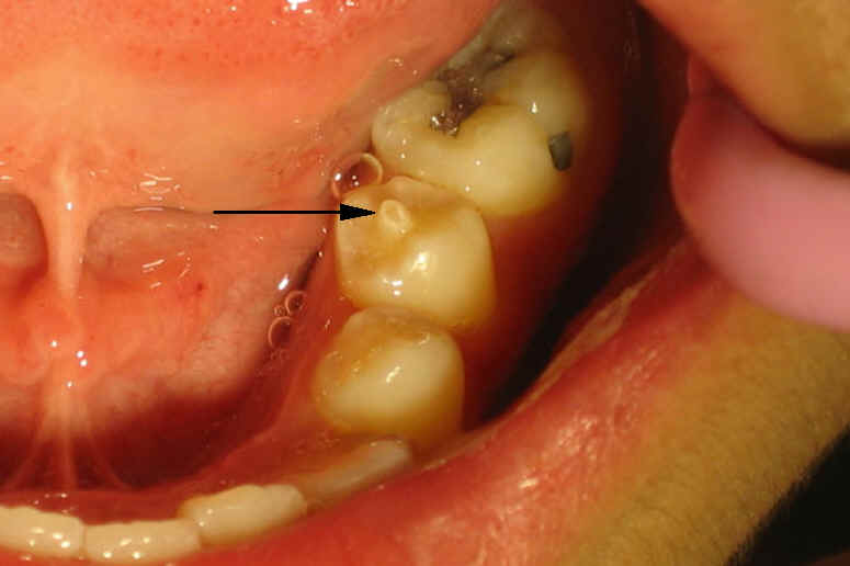
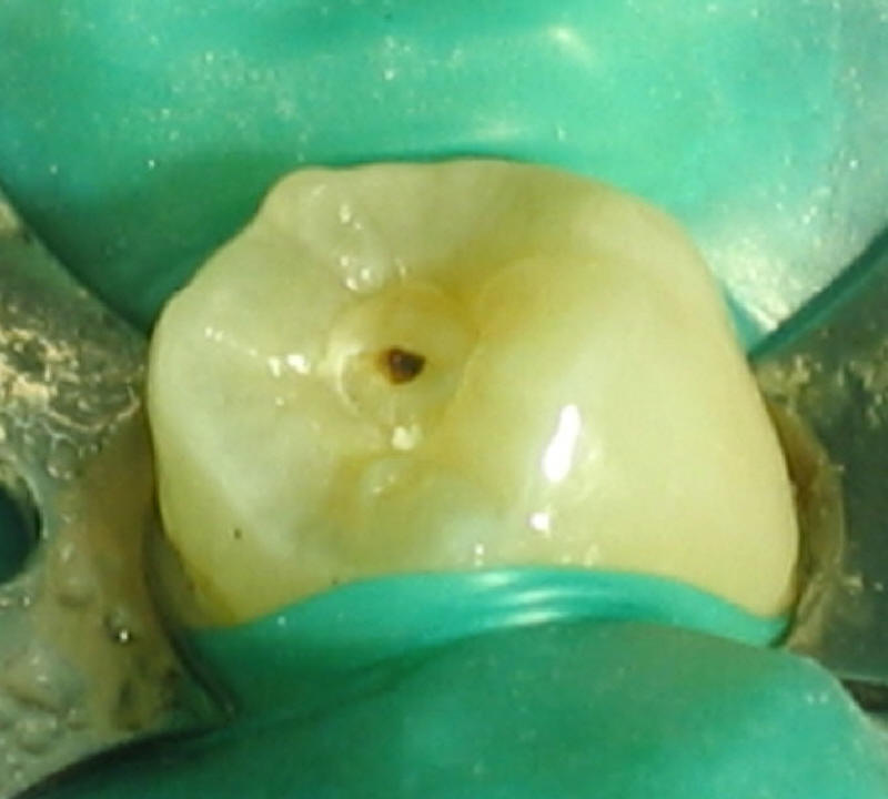
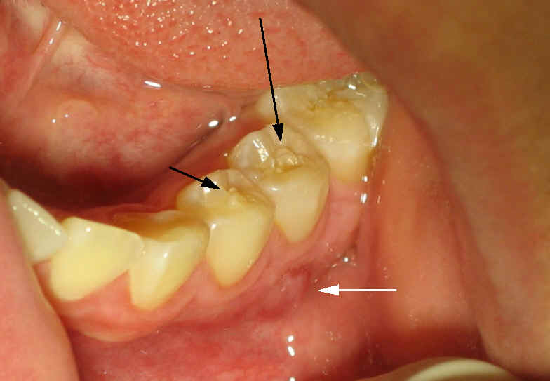
Central cusp is a type of tooth abnormality by birth and is most commonly found in Chinese descents. This abnormality is almost exclusively found in the premolars (black arrows in Fig.1 and 3). The long, slender cusp is easily broken, exposing the nerve (black dot in Fig2). The infected nerve causes an abscess at root tip (white arrow in Fig.3).
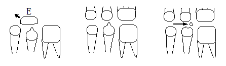
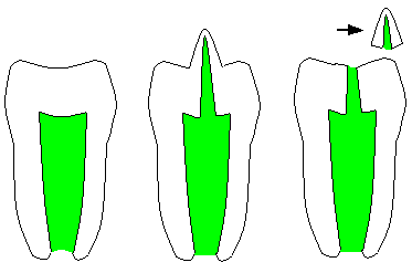
Let us use illustration to show you what happens to the central cusp. Fig.4 show that a premolar with a central cusp is replacing its predecessor, a baby molar tooth (E). Gradually the premolar is going to contact its opposing teeth (Fig.5). The sharp central cusp may break when we eat (arrow in Fig.6).
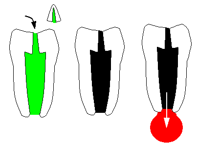
Fig.7 shows a cross section of a normal premolar with nerve in the middle (green). The nerve of the premolar with central cusp extends into the later (Fig.8). When the central cusp is fractured (arrow in Fig.9), the nerve is exposed (curved arrow in Fig.10). Very soon the exposed nerve is dying (black in Fig.11). The dead nerve causes serious root tip infection (red in Fig.12). In the next lecture, we are going to talk about how to treat root tip infection associated with a tooth with fractured central cusp. So long.
Xin Wei, DDS, PhD, MS 1st edition 05/21/2009, last revision 07/05/2009