|
|
|
|
| Fig.1 | Fig.2 | |
|
|
|
|
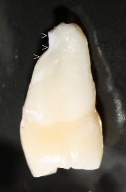
| Fig.3 | Fig.4 | Fig.5 |
Dental Education Lecture: CT before Braces
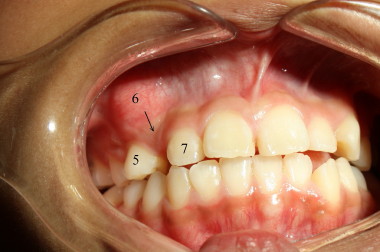
Tiffany is thirteen years old. Her teeth are pretty crowded (Fig.1). She needs braces. Upper right canine has not come out yet. There is a space between the teeth #5 and 7, where the canine (#6) should be found. In fact it is most likely above the tooth #5, underneath the gums. It looks like a big tumor.
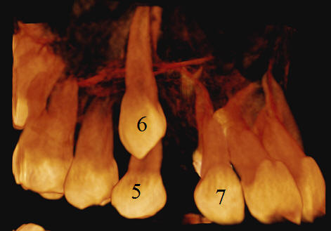
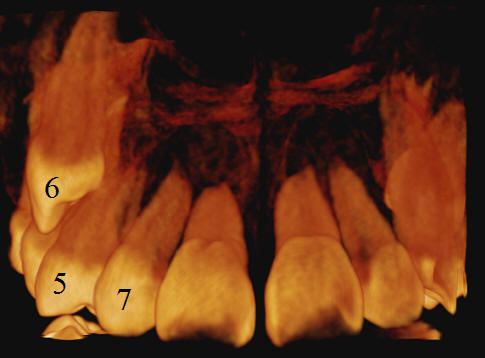
In order to arrive at correct diagnosis and treatment plan for braces, CT is taken. It shows that the tooth #6 is located above the root of the tooth #5 (Fig.2,3, three-dimensional images).
There are two types of treatment plans. The first one is to move the tooth #6 to where it belongs (as indicated by arrow in Fig.1). It is a lot of work. Besides, the space between the teeth #5 and 7 is narrower than the width of the tooth #6 (Fig.2). To increase the space for the canine, the front teeth may have to be pushed forward, creating a cosmetic problem.
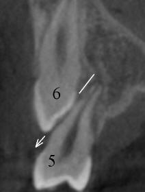
The second treatment plan is to extract the tooth #5 and let the tooth #6 erupt by itself. Why do we take out seemingly normal looking tooth #5 and keep unerupted tumor-like canine? The CT pictures (Fig.2,3,4 (two-dimensional)) demonstrate that the root of the canine is longer than that of the tooth #5.
In fact, the root of the tooth #5 is deformed, like a ditch, near the root tip (arrowheads in Fig.5) because of proximity of the tooth #6 against the root of the tooth #5 during their formation and eruption (arrow in Fig.4).
In brief, CT is an important diagnostic tool for complicated brace cases.
Xin Wei, DDS, PhD, MS 1st edition 04/15/2011, last revision 04/15/2011