|
|
|
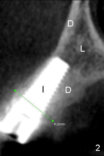
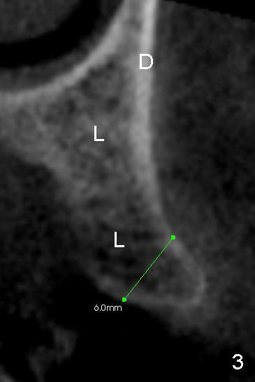
The cross section in Fig.2 (CT scan) shows that the width of bone holding the implant is 9 mm. The width of the bone before expansion is 6 mm, the same as shown in Fig.3. Fig.3 is the cross section of the other side (missing 2nd premolar). It appears that the bone is not expanded as much as expected (5+6=11 vs. 9 mm). In fact, the bone density next to the implant is increased (D (dense) in Fig.2, as compared to L (loose) in Fig.3 at the same level). So bone expansion is accompanied with bone compression/condensation. There is dense (hard) bone immediately around the implant using bone expansion. It makes the implant very sturdy in the bone. Return to main article
Xin Wei, DDS, PhD, MS 1st edition 10/12/2012, last revision 10/12/2012