|
|
|
|
|
|
|
|
|
|
|
Braces Before Implant: Design and Finish
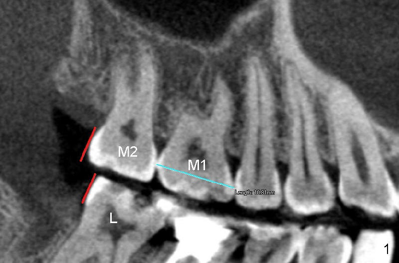
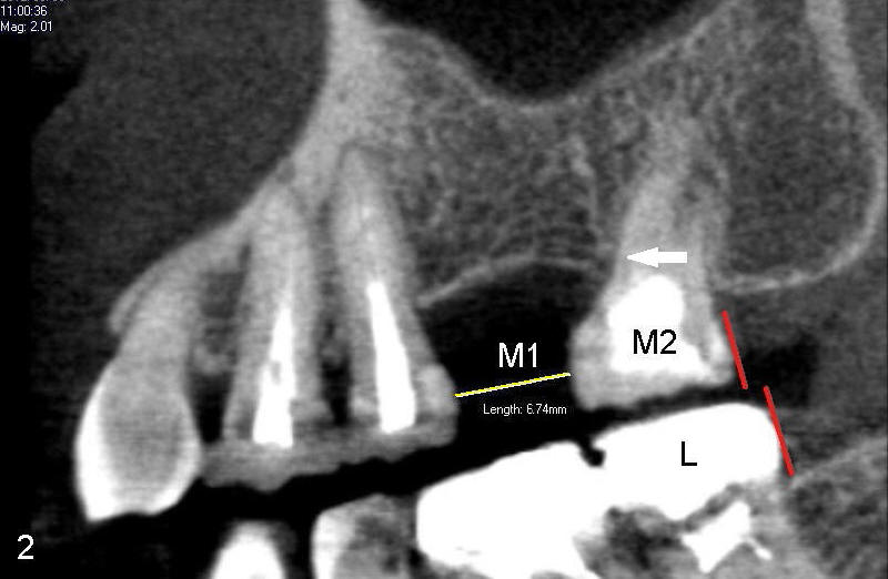
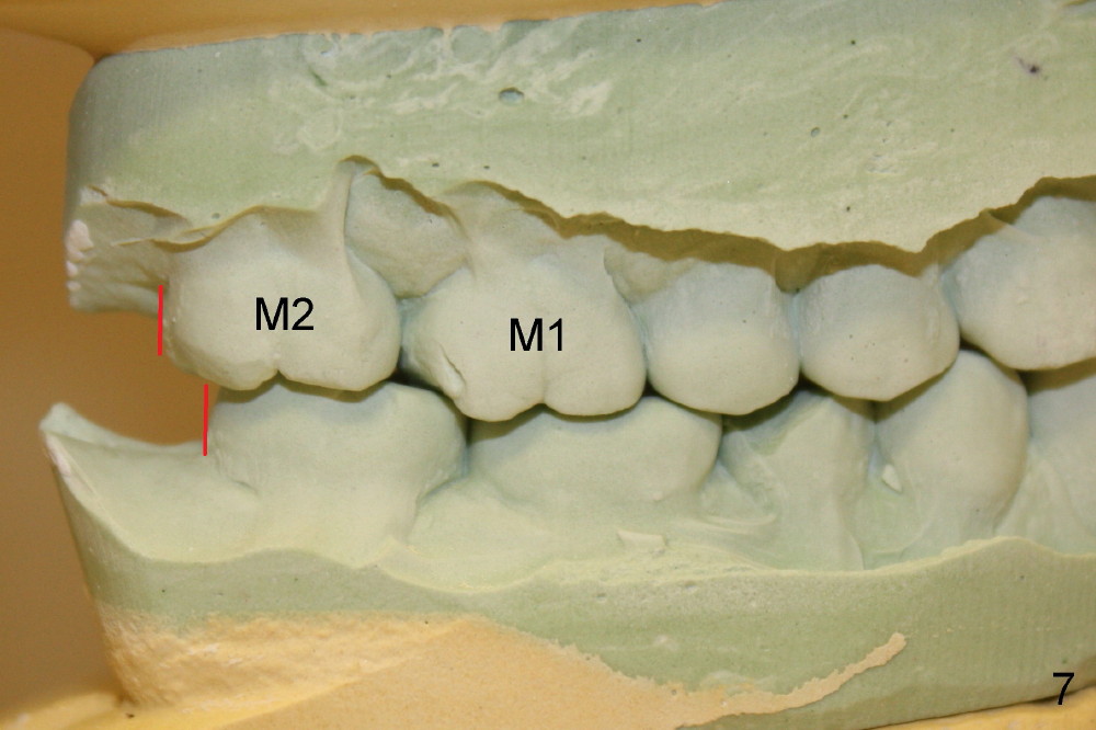
Ms. Li has lost upper left first molar (Fig.2 M1) for a while. Now she wants to have an implant, but the space is a little too small, as compared to that on the right (Fig.1 M1). The space decreases because the 2nd molar has shifted forward (Fig.2 arrow) since the first molar was extracted. Normally the upper 2nd molar is a little behind the lower one (compare Fig.1 M2 with L: red lines). By contrast, the left upper 2nd molar is in front of the lower one (Fig.2 M2 vs. L).
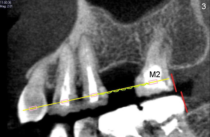
Before an implant can be placed for the missing molar, the space should be restored by pushing the 2nd molar backward. Three teeth in front of the missing molar gets braces (Fig.3 pink boxes). These three teeth act together as an anchorage to push the 2nd molar backward using spring (green coil) between them. A thicker wire is used to connect these four braces (yellow).
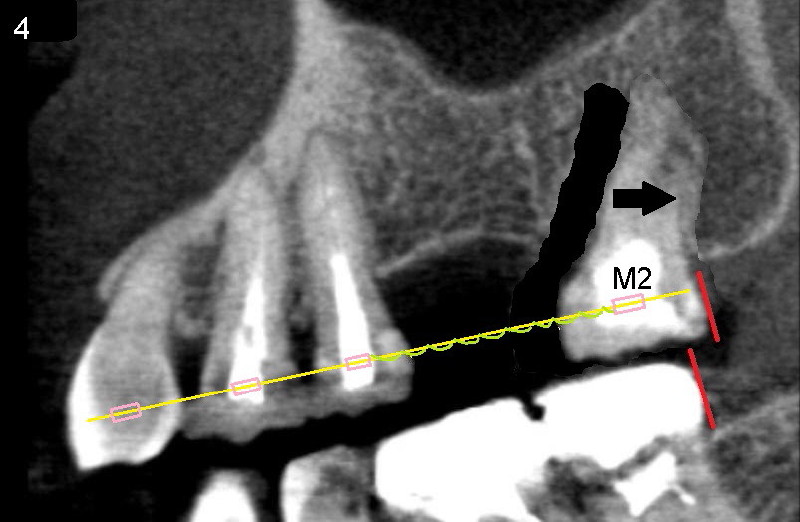
Under the tension of the spring (green), the 2nd molar will be moved to its original position in a few months (Fig.4 arrow).
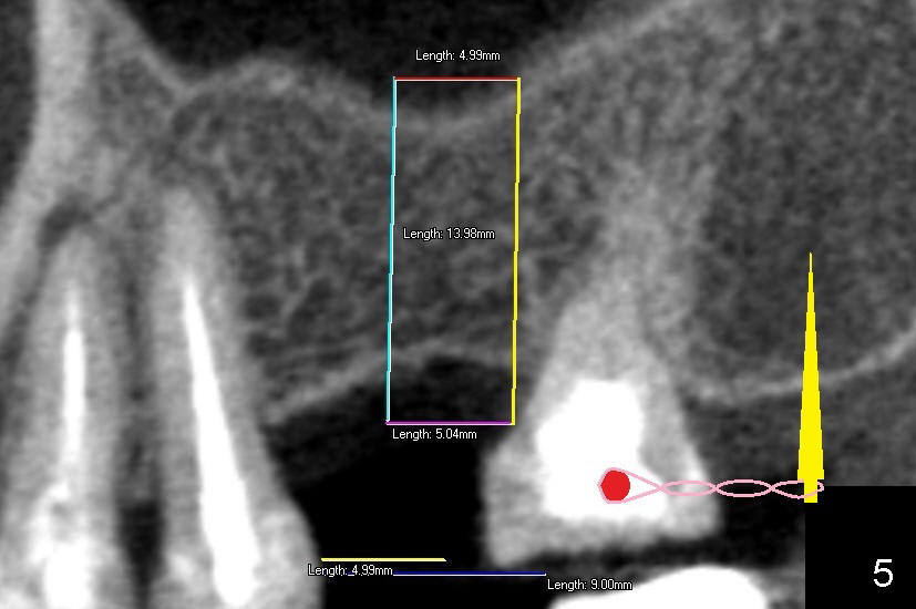
In fact, a mini-implant (Fig.5: yellow pin) is also used as an anchorage to move the 2nd molar backward under the tension of rubber bands (pink). The red circle in Fig.5 is a tiny brace to hold the other end of the rubber bands. Without enough space, it is difficult to place an implant (box in Fig.5).
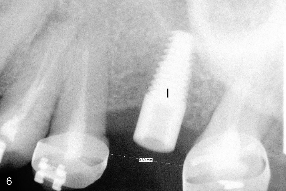
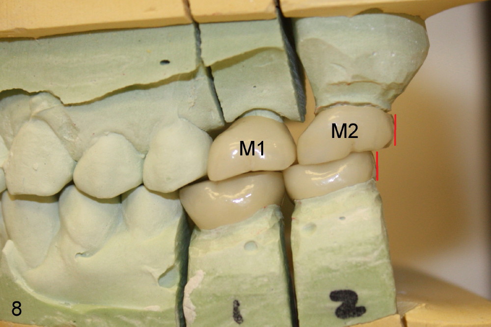
The space increases from 6.7 mm to 9.4 mm with local braces in 4 months (compare Fig.2 and Fig.6). An implant is placed (Fig.6: I: 5 mm in diameter, 14 mm in height). In 3 months, Ms. Li gets a new tooth (Fig.8 M1; four new crowns in dental model). Local bite relationship (red lines) is basically the same as the other side (Fig.7). We wish her the best.
Xin Wei, DDS, PhD, MS 1st edition 05/23/2012, last revision 07/09/2020