 |
|
Dental Education Lecture: Changes after Extraction
Sometimes we have a bad tooth with severe pain. After discussion, we decide to get it out. Pain is gone with it. We feel everything alright and ignore the fact that we need to return to dental office for further treatment. Why bother when we are pain-free?
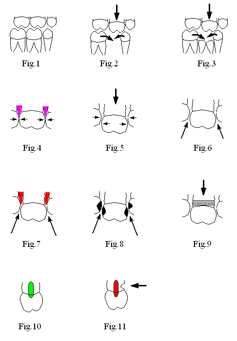
Fig.1 shows six normal back teeth. A little while after we remove the middle molar tooth on the bottom, the adjacent bottom teeth begin to shift and tilt toward the missing tooth area (two curved arrows), while the upper opposing tooth moves downward (straight arrow). Every change occurs gradually, painlessly and unnoticeably. Interestingly, some of my patients have a severely broken down tooth with remaining root tip. They are reluctant to have it out. Their reason is that the root tip can hold the space. In fact, it is the crown of a tooth that holds the space, but not the root. Fig.3 shows that the crown of the middle bottom tooth is gone. Similar changes (shifting, tilting and elongation) happen, like those in Fig.2. These positional changes make local oral hygiene difficult to maintain.
Fig.4 shows normal contact among 3 upper teeth (indicated by arrows). The pink areas in Fig.4 represent normal gums between the teeth. When the middle tooth starts to shift downward (big arrow in Fig.5), the normal contact between the teeth is lost. Food easily gets into there (arrows, Fig.6). Gums get irritated (red, Fig.7) and cavities develop in the areas between the teeth (black, Fig.8) due to food impaction and retention of germs there.
When the middle tooth drops down, the gums may not come down as much. The root gets exposed. Improper brushing creates a notch in the cervical (neck) area between the crown and root (Fig.9). Since there is no enamel in the cervical area, it gets wear and tear easily. Let turn the middle tooth in Fig.9 for 90 degrees. The cervical notch is pointed by arrow. The bottom of the notch is close to the nerve (red). When you brush, toothbrush bristles enter the bottom of the notch, causing tooth sensitivity. Compare it with Fig.10 showing a normal tooth without a cervical notch and with normal nerve (green). If you like to know how to treat the cervical notch, look at Fig.3 and 4 in lecture Cases of Filling.
Changes after extraction mentioned above happen slowly without your noticing. The sooner we restore the missing tooth, the less severe complications we are going to have. Next lecture we are going to discuss how to fix these complications in great length.
Xin Wei, DDS, PhD, MS 1st edition 02/14/2009, last revision 02/15/2009