
|
|
|
|
|
| Fig.1 | Fig.2 | Fig.3 | Fig.4 |
|
|
|
|
|
| Fig.5 | Fig.6 | Fig.7 | Fig.8 |
|
|
|
||
| Fig.9 | Fig.10 | ||
Dental Education Lecture: Bone Graft Before Implant
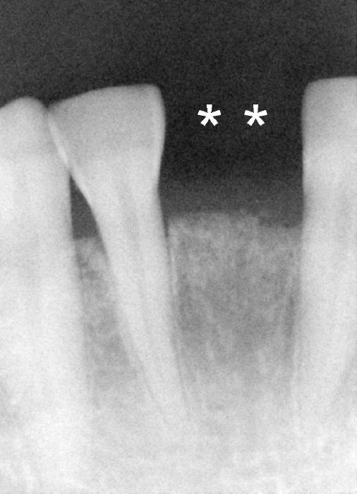
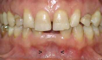
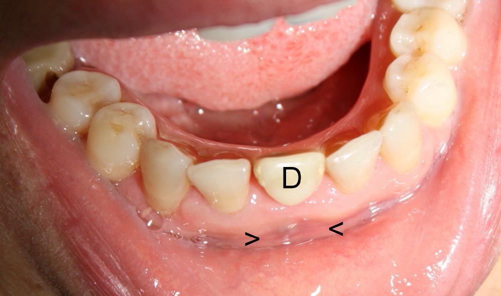
Ms. Yang does not have two adult incisors on the bottom (Fig.1 (**), and Fig.2). When the baby teeth fall out in her late twenties, she has to wear a denture (D in Fig.4). She cannot eat with the denture. The denture is so loose that it may shoot out of her mouth like an arrow when she talks. By the way, Ms. Yang is an elementary school teacher. She does not want to embarrass her students with her loose denture. Finally we decide that she needs an implant.
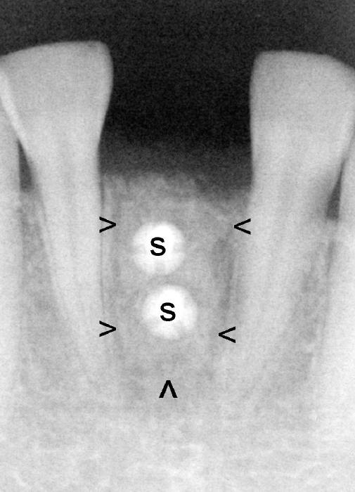
However, the bone to hold the implant is so skinny (sunken, arrowheads in Fig.2) that we have to make this area big and thick by placing a piece of bone (arrowheads in Fig.3) there, fixed with two tinny tiny screws (S, Fig.3). Fig.4 arrowheads indicate that the area is much improved as compared to Fig.2.
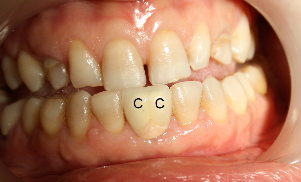
Four months after bone graft, two tiny screws are removed and an implant (I in Fig. 5) is placed. Another four months later, an abutment (A in Fig.5) is installed and finally two joined crowns are cemented (C in Fig. 5,6). Now Ms. Yang has two beloved baby teeth back. They are very stable. She is not going to have nightmare that the teeth fall in front of her students or colleagues.
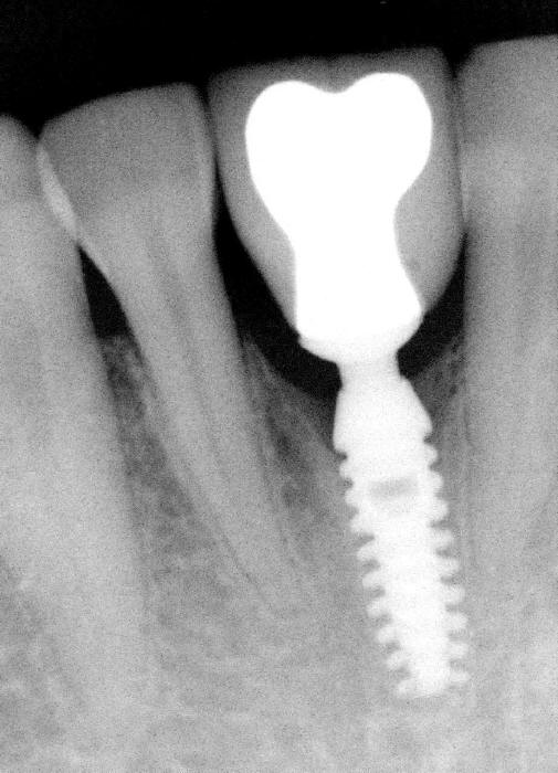
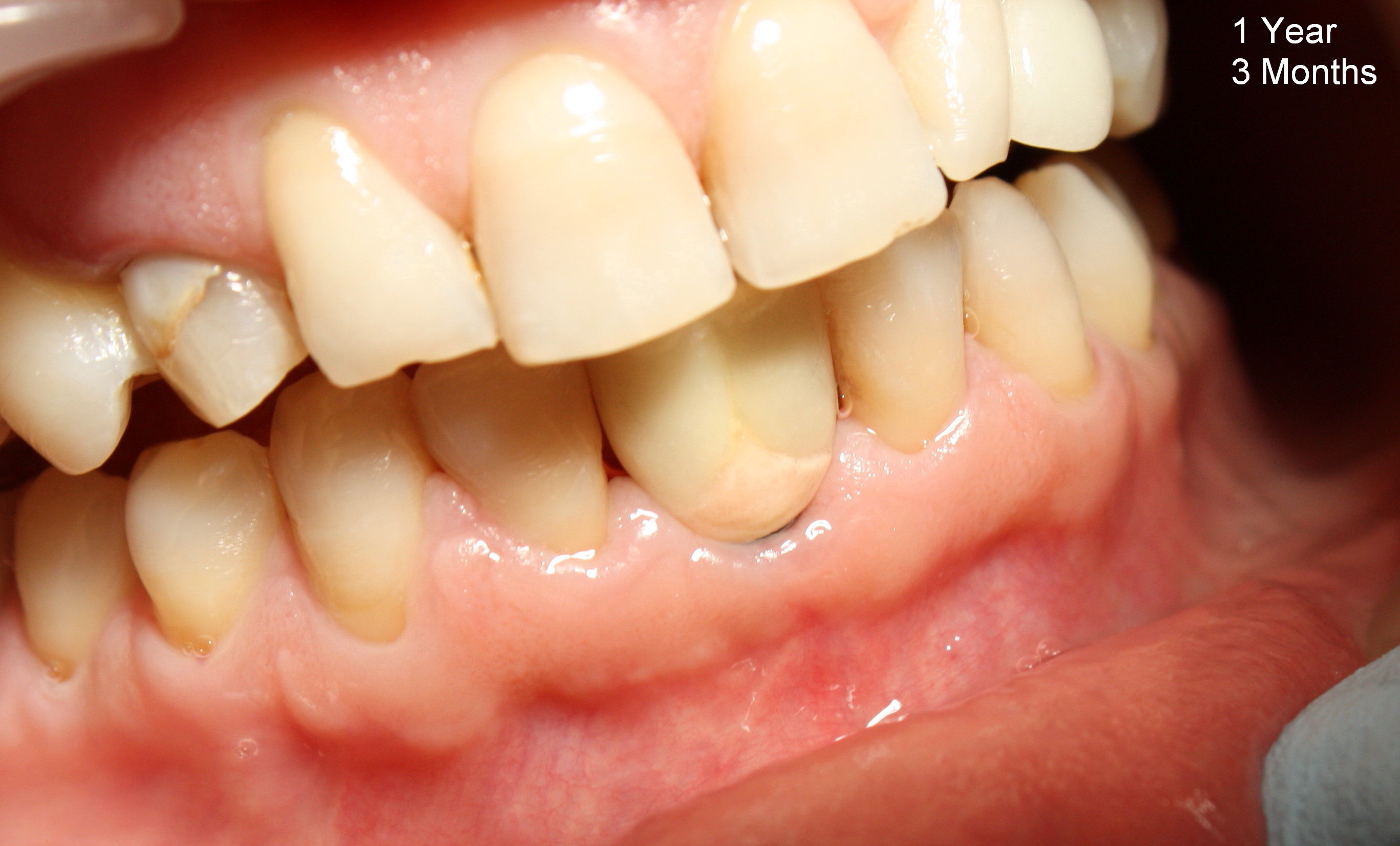
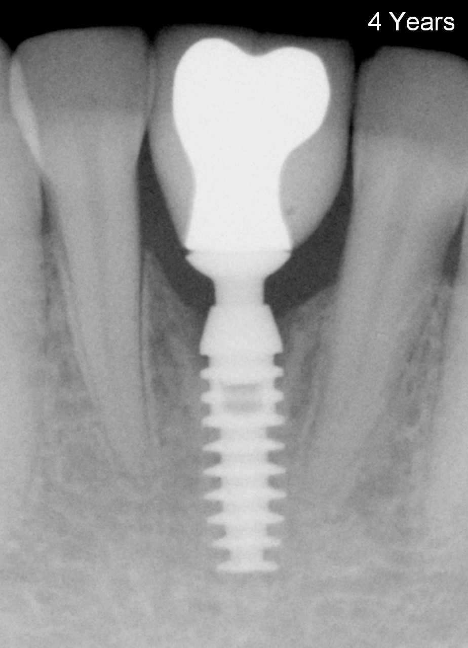
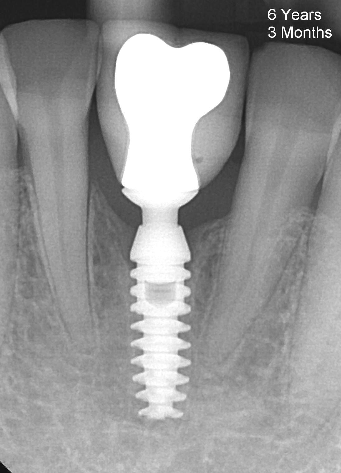
Bone remains stable around the implant 2 years after crown cementation (Fig.7). The gums around the crowns looks healthy (Fig.8). The patient eats normally. The implant remains solid 4 years (Fig.9) and 6 years (Fig.10) after cementation.
Xin Wei, DDS, PhD, MS 1st edition 06/25/2010, last revision 12/15/2016