
|
|
|
|
|
| Fig.1 | Fig.2 | Fig.3 | Fig.4 |
|
|
|
|
|
| Fig.5 | Fig.6 | Fig.7 | Fig.8 |
|
|
|
||
| Fig.9 | Fig.10 | ||
Chin Graft for Lower Incisors
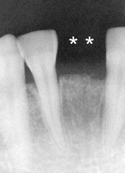
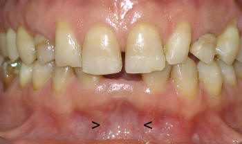
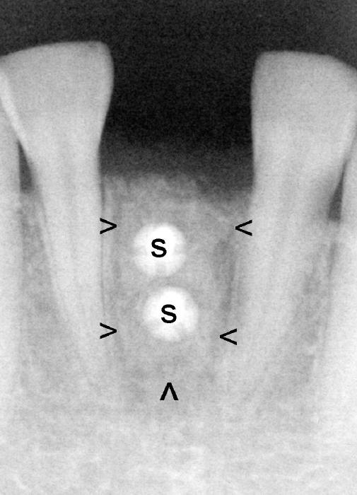
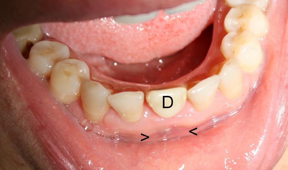
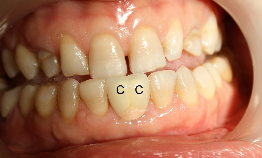
Two mandibular central incisors are congenitally missing from a healthy 36-year-old lady (Fig.1 (**), and Fig.2). The ridge is atrophic, especially apical to the crest (Fig.2 arrowheads). A corticocancellous block is harvested from the chin and fixed in place with 2 screws (Fig.3 S). The patient is doing well postop probably due to minimal invasive approach (single donor/recipient site). The buccal plate morphology improves postop (Fig.4 between arrowhead, apical to a removable denture (D)). Five months later, a 3.5x11 mm Bicon implant is placed. Two "mini" crowns with pink gingiva are cemented 8 months later (Fig.5,6).
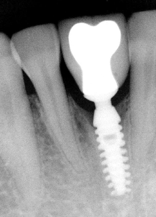
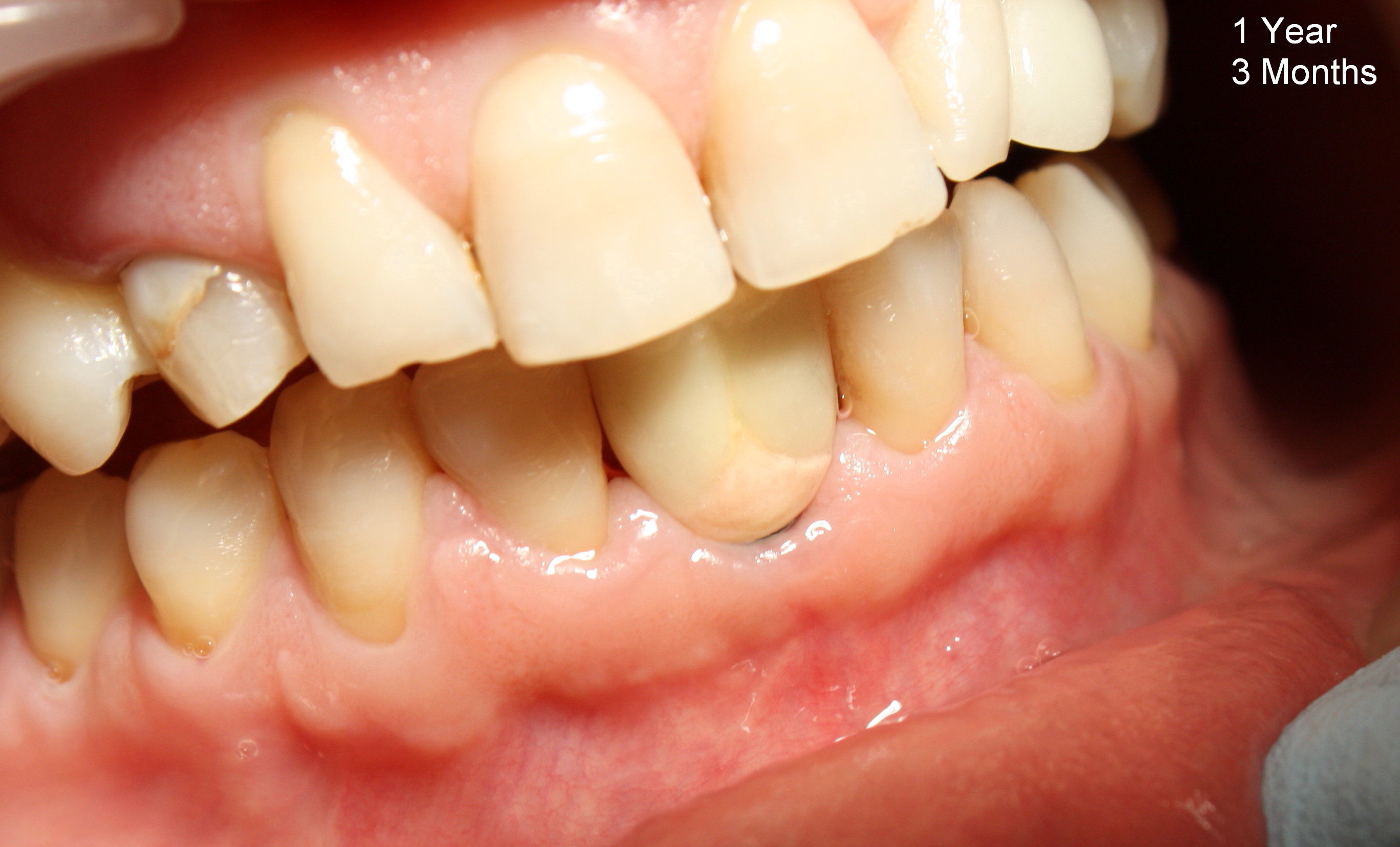
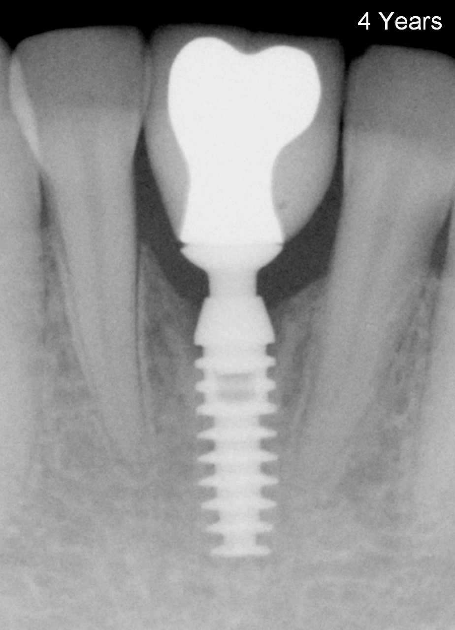
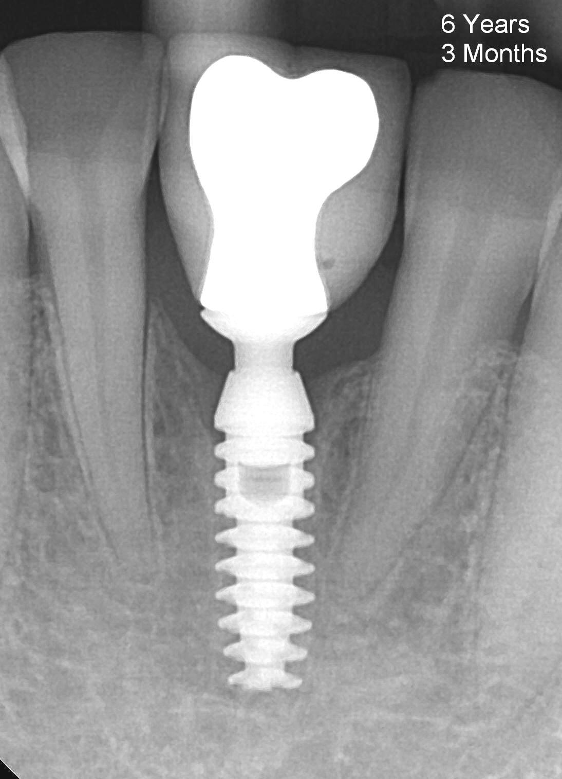
Crest bone remains stable around the implant 1 year 3 months after crown cementation (Fig.7). The gingiva is healthy (Fig.8). Recently the patient returns for follow up There is no bone loss 4 years (Fig.9) or 6 years 3 months (Fig.10) post cementation. She eats normally and is pleased with the result.
Return to Fellowship Documentation Form, Lower Incisor Immediate Implant
Xin Wei, DDS, PhD, MS 1st edition 06/25/2010, last revision 12/15/2016