
|
|
|
| Fig.1 | Fig.2 |
|
|
|
| Fig.3 | Fig.4 |
Dental Education Lecture: Gum Recession after Extraction
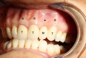
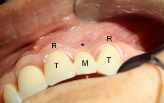
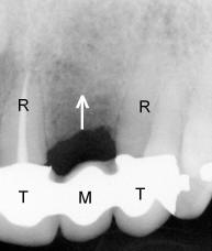
Mrs. Yang has a beautiful bridge (TTMT in Fig.1) to replace a missing tooth (M) in the front. She can chew very well, but she is not happy with the fact that the portion of the fake tooth near the gum line (* in Fig.2) is not covered by the gums (G). This is a cosmetic issue. Closer look of the missing tooth area shows that there is a big crater-like defect (concavity) over the root portion of the missing tooth (* in Fig.1, 3). By contrast, the roots of the natural teeth (R) are bulging (convex), suggesting that after extraction both soft (gums) and hard (bone) tissues are shrinking (in depth). By the way, the tooth was extracted 7 years ago. X-ray in Fig.4 shows that the bone in the missing tooth area also shrinks in the vertical dimension (arrow).
To solve Mrs. Yang's gum issue, we need to put back not only gum tissue, but also bone. However, the gums and bone will shrink again over the years. Because without a root, the gums and particularly the bone have no functioning stimulus. Therefore, after replenishing the soft and hard tissues, we need to place an implant for the missing tooth for long-term cosmetic effect.
This case tells us a good lesson. After extraction, we should have an implant as soon as possible to prevent no-use shrinking. Implant replaces our missing root. With root or implant, our bone has normal functioning. Although bridge and partial denture can restore our chewing ability, they do not prevent gum and bone shrinking, because they do not replace our missing root. In all, implant is the best solution to single or several missing teeth. Shrinkage can happen very quickly after extraction. Therefore after extraction, we need to return to dental office to have an implant placed as soon as possible.
Xin Wei, DDS, PhD, MS 1st edition 05/09/2010, last revision 09/29/2012