|
|
|
|
|
|
|
|
|
|
|
|
Socket Collapses After Extraction
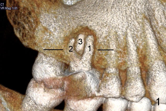
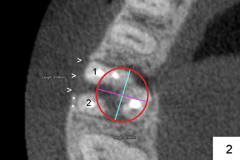
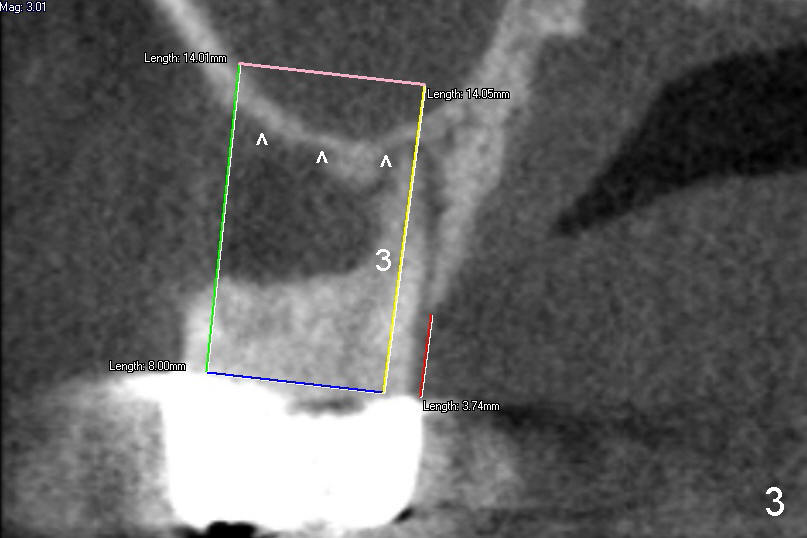
Six months ago, an article was written about one of John's upper molars. It had so severe infection that it would be extracted. There was severe bone loss (a big hole) around its three roots (Fig.1 (CT 3-dimensional image): 1,2,3). It was planned to place an implant about six weeks after extraction. At that time, the socket was wide and long. A wide and long implant was planned (Fig.2, 3, 4: 5 mm in diameter and 14 or 17 mm in length).
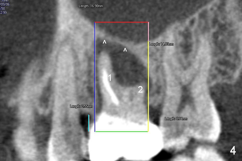
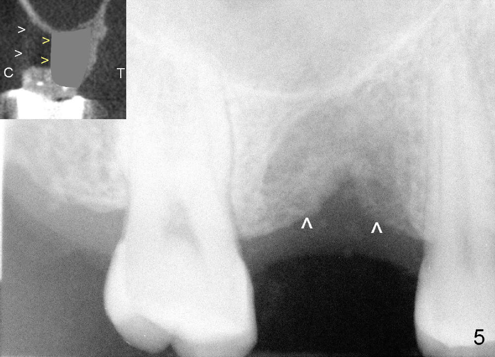
After extraction, preauthorization was submitted to his dental insurance. It took six months (instead of six weeks) to get approval. John returns for implant. But the socket shrinks substantially in up and down direction (Fig.5 arrowheads, as compared to Fig.1). The socket collapses more in cheek-to-tongue direction (C, T), i.e., from white > to yellow ones (Fig.5 insert), as compared to Fig.3).
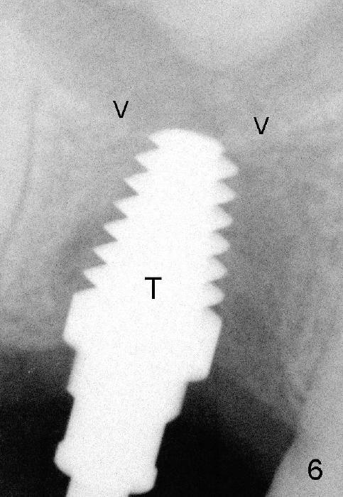
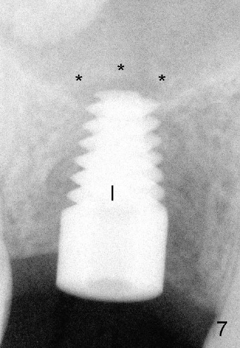
A much smaller (6 mm) and shorter (11 mm) implant is placed (Fig.7 I) after using a tap (Fig.6 T) to form threads in the bone. Drills and tap have to gently break the sinus floor (Fig.6 arrowheads) into the sinus so that the top end of the implant can be engaged into the tough portion of the bone for stability. The sinus floor has been raised like a rainbow (Fig.7 ***).
In all, an implant should be placed immediately or soon after extraction.
Xin Wei, DDS, PhD, MS 1st edition 09/09/2012, last revision 03/31/2013