 |
|
Dental Education Lecture: Tooth Wear and Tear
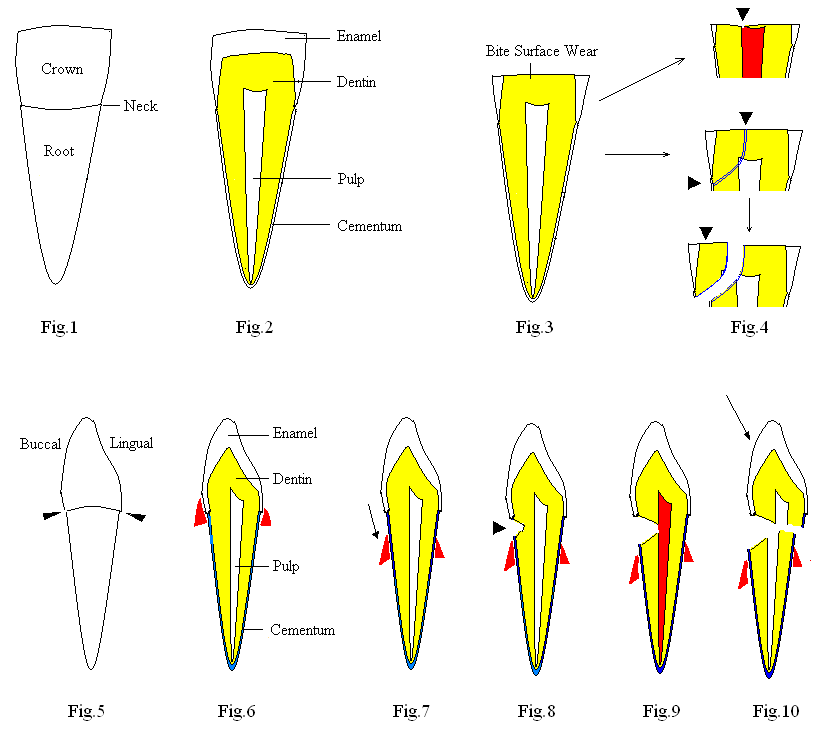
There are two common types of tooth wear and tear: one in the biting surface and the other one in the cervical area (neck of the tooth). We should review tooth anatomy before discussion of these abnormalities.
Fig.1 shows the front view of a lower incisor. It has the crown, root and neck. Fig.2 is a cross section of Fig.1, revealing three hard tooth structures: enamel, dentin (yellow) and cementum and one soft tissue: pulp or nerve. The enamel covers the crown, whereas the cementum covers the root. As we know, the enamel is the hardest tissue in our body, resistant to wear and tear. However, sometimes due to our bad habits: such as chewing ice and grinding teeth at night, the enamel in the biting surface may be worn prematurely (Fig.3). You can go to When We Need Crown lecture and look at Fig.3 to see what biting surface wear looks like in a molar tooth. Continuous wear and tear leads to exposure of the nerve (arrowhead, top drawing in Fig.4) and causes nerve infection and pain (red). Severe wear and tear weakens the tooth and there is a cracking line over a portion of the tooth (indicated by arrowheads in the middle drawing of Fig.4). When you accidentally bite on a piece of sand, the tooth is broken (bottom of Fig.4). If the break line is close to the nerve or across it, pain is excruciating. Wearing a night guard is the most effective way to prevent excessive occlusal surface wear and tear.
The second common type of tooth wear and tear is found in the neck of the tooth. Let us turn the tooth in Fig.1 for 90 degrees so that the buccal surface of the tooth (next to our lip) is on your left and the lingual surface (next to our tongue is on your right (Fig.5). Arrowheads point to the neck of the tooth. Fig.6 is a cross section of Fig.5, showing the inner structures of the tooth. The labeling is the same as that in Fig.2 except the cementum being painted blue. We also add the gums (red) to the illustration at the neighborhood of the neck. If you ignore oral hygiene for a long time, your gums are receded (as shown by arrow in Fig.7). Probably due to improper brushing, a notch is formed in the neck of the tooth (arrowhead, Fig.8). Deepening of the notch results in nerve exposure and severe pain (red, Fig.9). Notching also weakens the tooth and sudden biting force (arrow in Fig.10, such as biting upon chicken or fish bone) can cause tooth fracture. An early sign of a notch in the cervical area is tooth sensitivity while you are brushing, corresponding to the shallow notch shown in Fig.8. It is best to get it filled with white filling before it is too late. See Fig.3 and 4 in Cases of Fillings lecture.
Xin Wei, DDS, PhD, MS 1st edition 02/15/2009, last revision 02/15/2009