 Fig.3
As mentioned before,
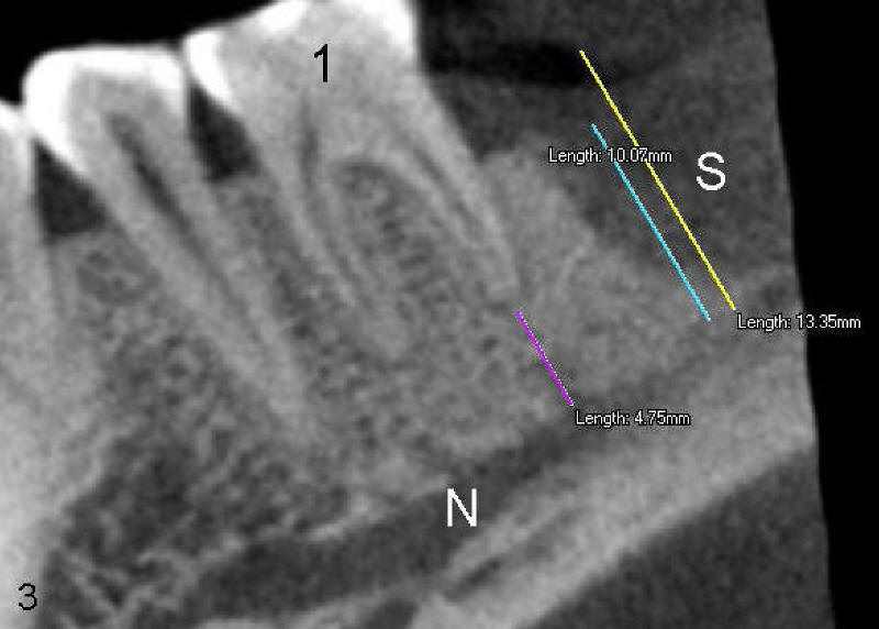
routine dental X-ray does not show the nerve precisely. Two months after
extraction of the 2nd molar, oral CT is taken. It allows the doctor to do
various measurements from the nerve (N) to the gums (yellow line), to the top of
bone (blue) and to the root tip of the neighboring tooth (purlple). At
that time, the socket (S) is healing, back to
original article
Fig.3
As mentioned before,
routine dental X-ray does not show the nerve precisely. Two months after
extraction of the 2nd molar, oral CT is taken. It allows the doctor to do
various measurements from the nerve (N) to the gums (yellow line), to the top of
bone (blue) and to the root tip of the neighboring tooth (purlple). At
that time, the socket (S) is healing, back to
original article