|
|
|
|
|
|
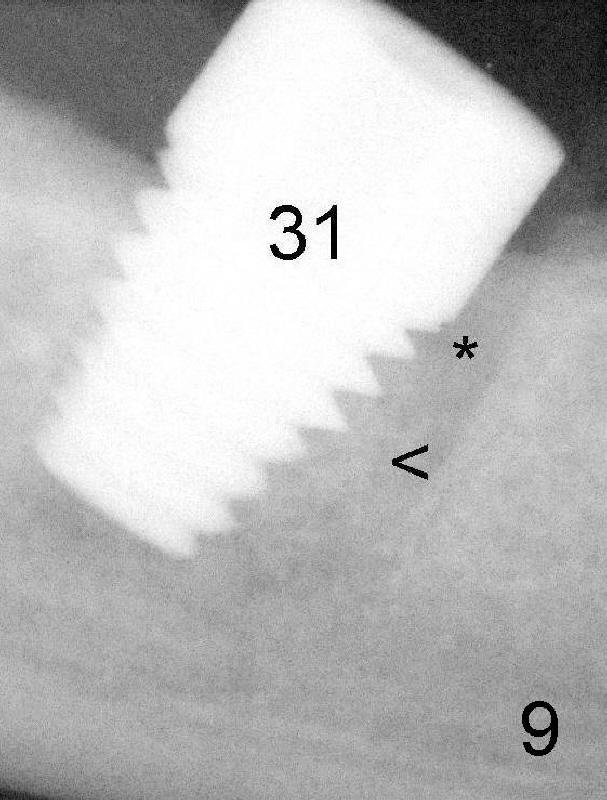
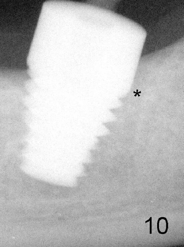
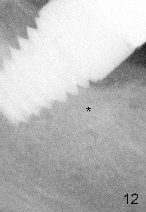
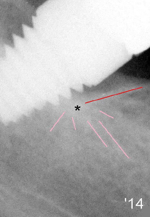
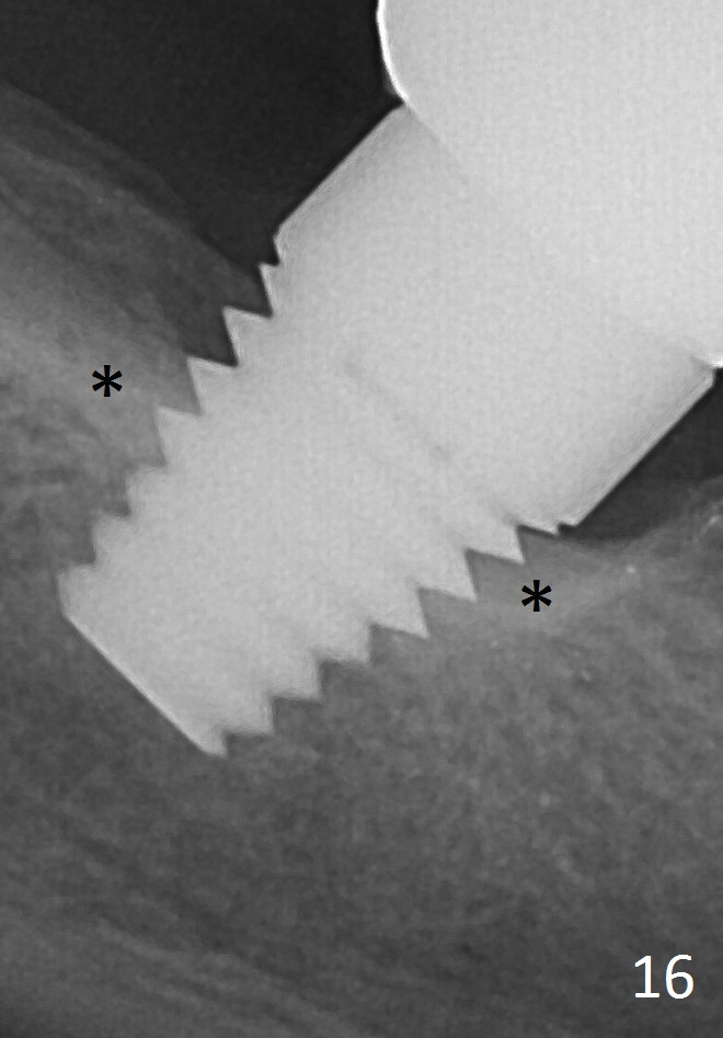
Regular X-ray in Fig.9 shows that the implant is just placed at the site of #31. Bone (<) is compressed into the bottom half of the mesial socket (*). Four and a half months later, the top half of the mesial socket is filled by new bone (Fig.10: *). Another three months the bone density in the previous mesial socket (Fig.12 *) is as high as that of the neighboring bone. One year 9 months after crown cementation, bone next to the implant at the site of #31 has radiating pattern (Fig.14), suggesting that the implant sustains a lot of chewing force. The bone is denser around the implant 4 years after crown cementation (Fig.16 *).
Return to main article
Xin Wei, DDS, PhD, MS 1st edition 01/20/2013, last revision 05/04/2017