|
|
|
|
|
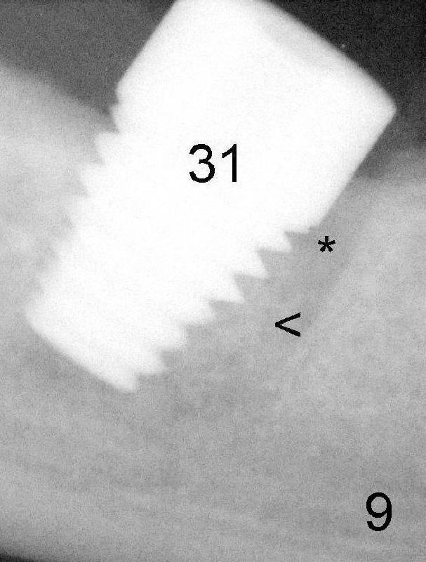
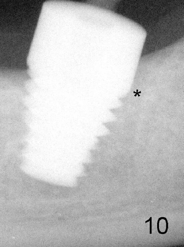
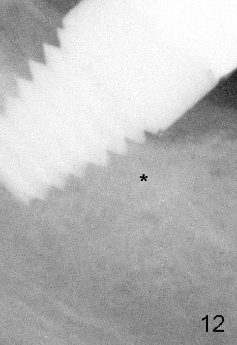
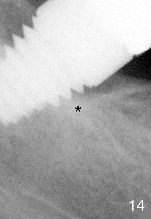
PA in Fig.9 shows that the implant is just placed at the site of #31. The septal bone (<) is compressed into the lower half of the mesial socket (*). Four and a half months later, the upper half of the mesial socket is filled by new bone (Fig.10: *). Another three months (after cementation) the bone density in the previous mesial socket (Fig.12 *) is as high as that of the neighboring bone. One year 9 months post cementation, bone trabecules have radiating patterns from * (Fig.14).
Return to Immediate Implant in 2nd Lower Molar
Xin Wei, DDS, PhD, MS 1st edition 01/20/2013, last revision 11/01/2014