|
|
|
|
|
|
Dental Education Lecture:
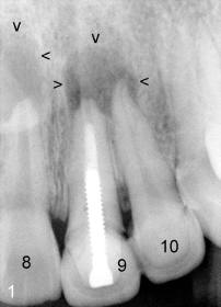
Fig.1 shows large radiolucent lesions associated with the teeth #8-10 (arrowheads). Treatment plan is to remove #9,10 crowns (joined), remove the post from #9, redo root canal therapy (RCT) and RCT for #8 and 10.
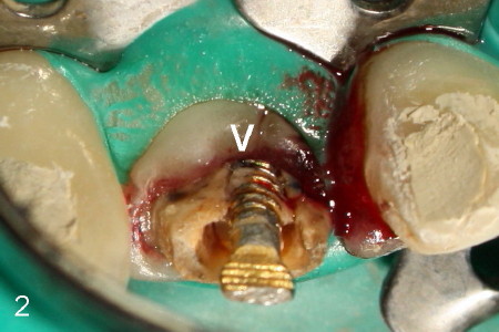
After #9,10 crowns' removal, subgingival margin was found in the lingual of #9 (Fig.2: arrowhead). It appears necessary to extrude #9 before restoration with single unit crown. Initially it was difficult to remove the post. RCT started first for #8 and 10.
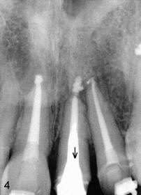
Fig.3 demonstrates initial bracketing. Note the uneven bracket placement in anterior segment for extrusion's purpose.
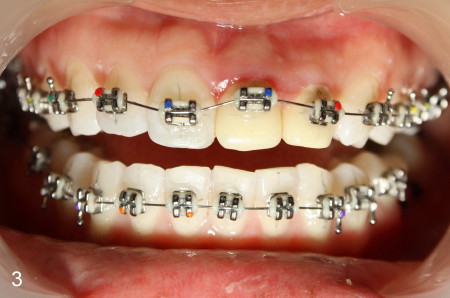
PA taken 8 months after orthodontic treatment shows that the tooth #9 has been extruded (Fig.4 arrow) and periapical radiolucency disappears.
A shortcoming of orthodontic extrusion in this case is that the gingiva is also extruded. Gingivectomy has been done several occasions for cosmetics. It appears that crown lengthening is necessary for long term effect.
Xin Wei, DDS, PhD, MS 1st edition 05/072011, last revision 03/23/2018