|
|
|
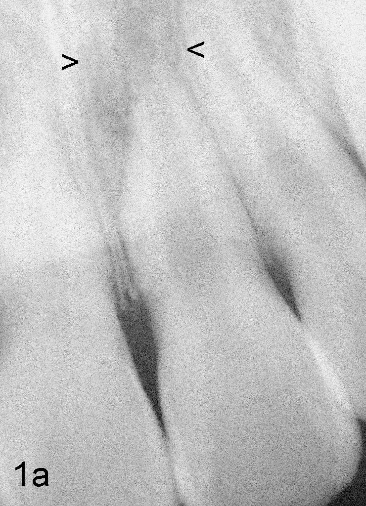
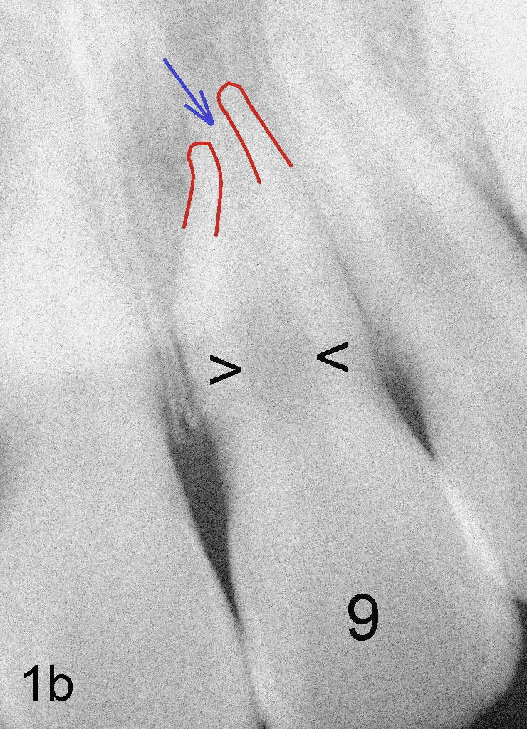
Fig.1 An upper central incisor (#9) before (1a) and after (1b)
drawing to show an enlarged canal (between black arrowheads in 1b) and open apex
(blue arrow). Arrowheads in 1a indicate a periapical radiolucent lesion
due to necrotic (died) pulp one year after trauma in a 10-year-old girl.
Return to main article or
Assistant Page
Xin Wei, DDS, PhD, MS 1st edition 07/12/2011, last revision 07/12/2011