

|
|
|
|
|
|
|
|
|
|
|
|
|
|
|
|
|
|
|
|
|
|
|
|
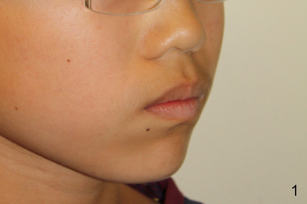
Extraction Ortho for Maxillary Protrusion
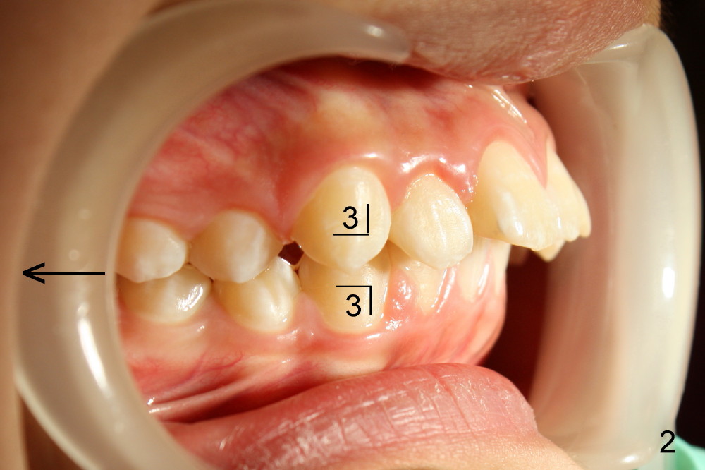
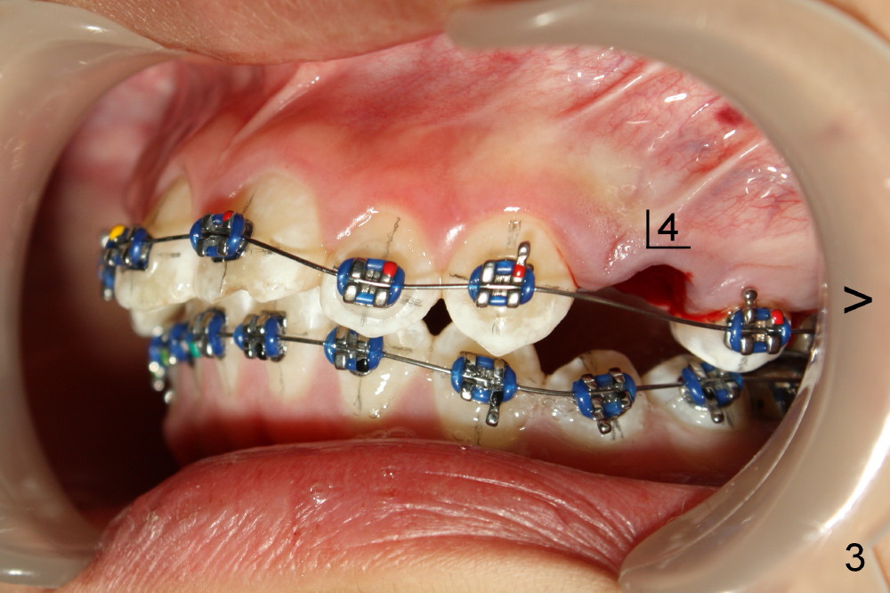
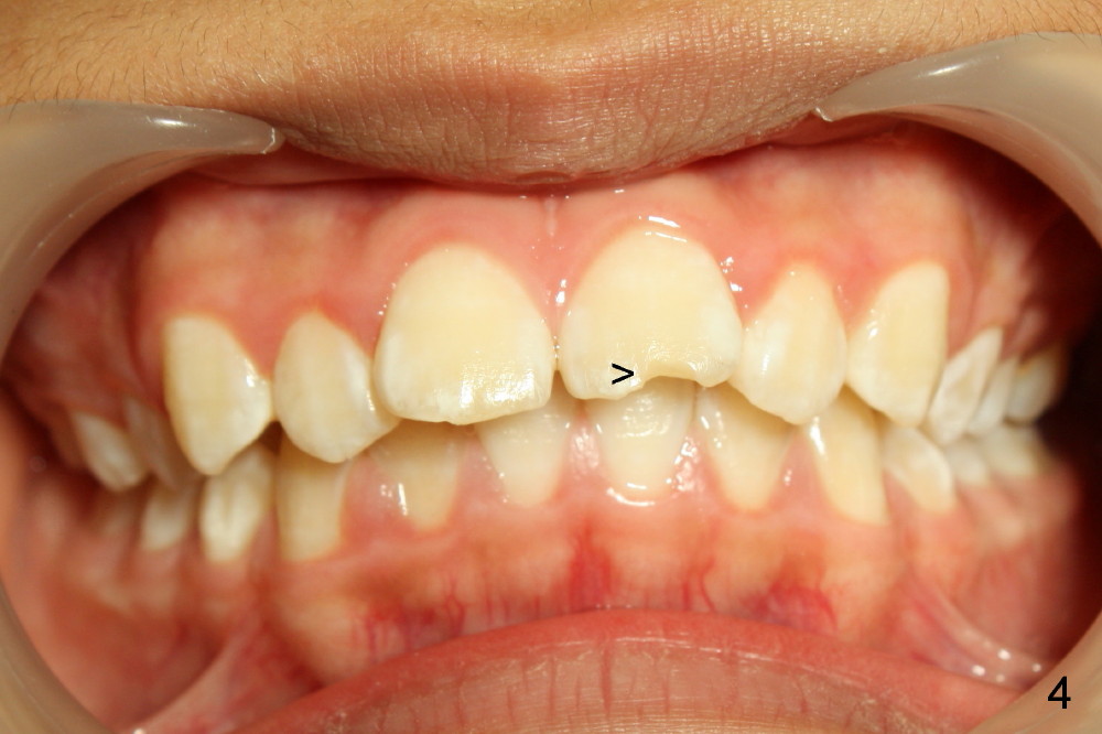
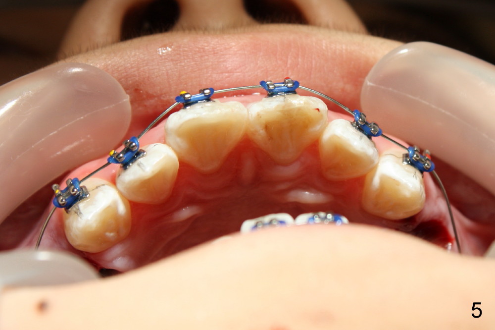
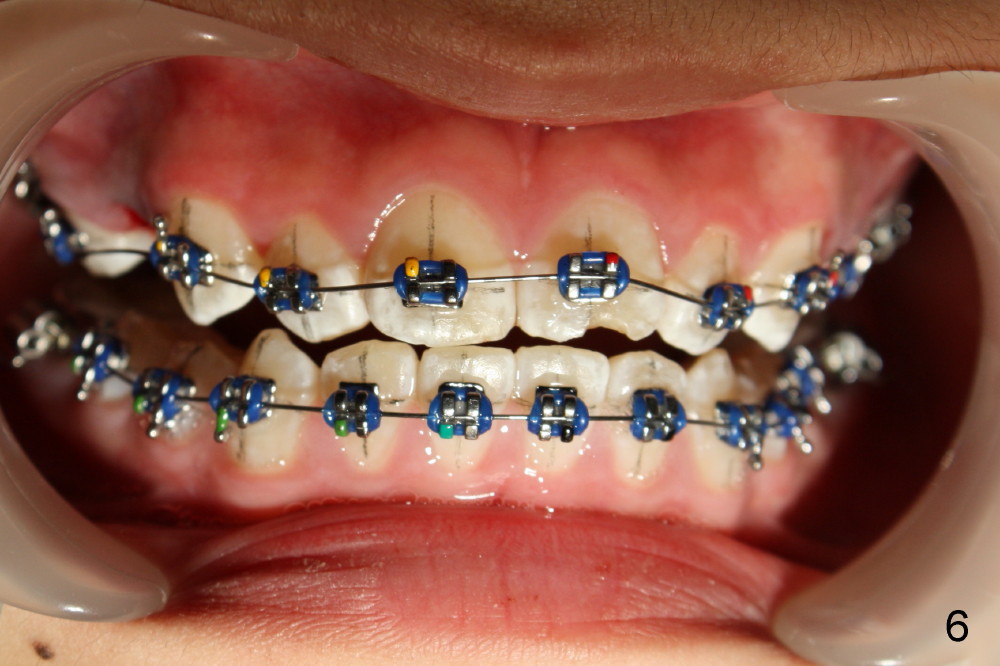
A 12-year-old boy has upper anterior protrusion (Fig.1-3). It is characterized with canine Class II malocclusion (Fig.2,3) and relatively wide crowns of these anteriors (Fig.4). It appears that one of the most efficient treatment modalities is orthodontics with extraction of the upper 1st bicuspids (Fig.3,5,6, photos taken immediately post extraction).
The 1st stage of treatment is alignment of the arches and correction of deep overbite (Fig.2-4,6) so that the upper anteriors have freedom to be distalized.
The 2nd stage is to retract the anteriors, either by power chains 6 to 6 or retract 3s, followed by incisors, according to occlusion at the conclusion of alignment.
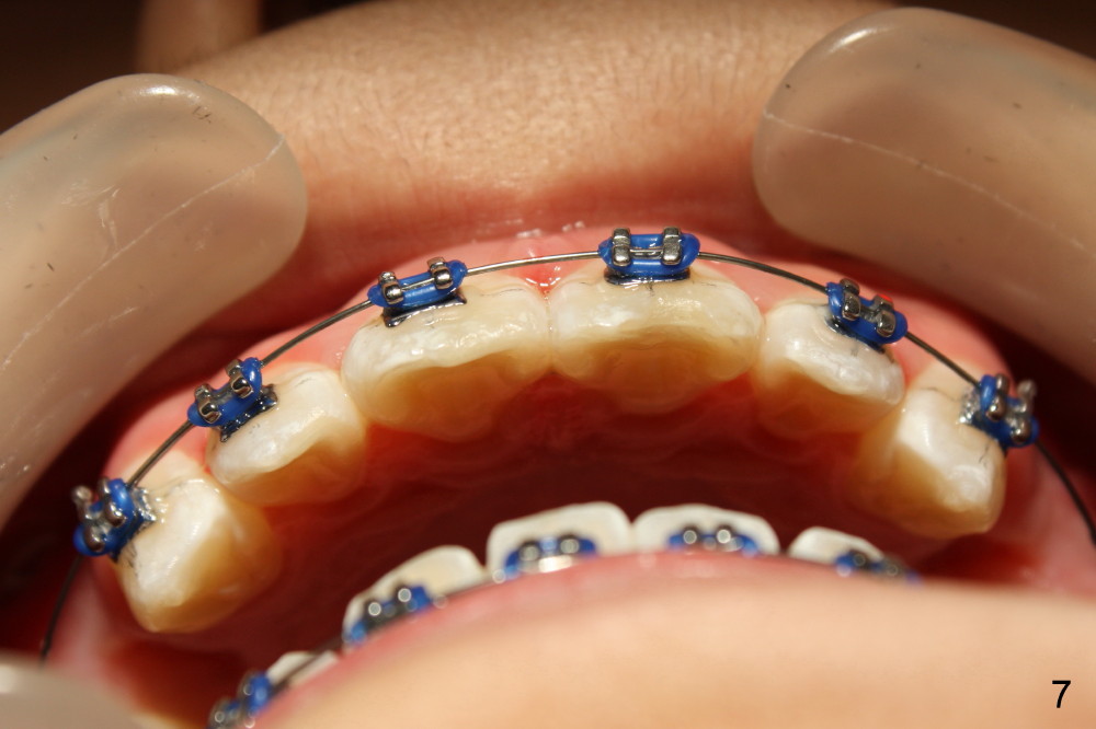
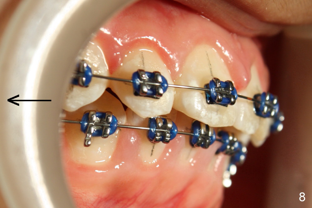
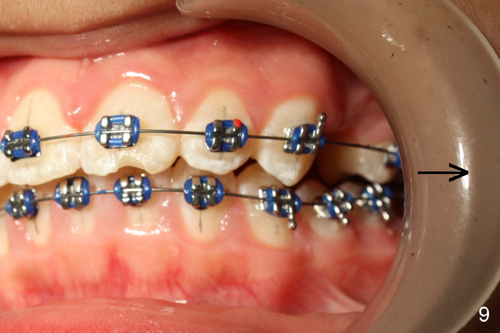
Fig.7-9 are taken 1 month post-bracketing. Alignment of the upper anteriors improves.
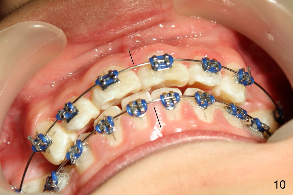
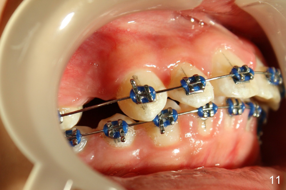
Fig.10-12 are taken 2 months post-banding. There is not enough clearance between the upper and lower incisors to retract the upper ones (Fig.10). At the present moment, the lower incisors have not been intruded, as indicated the upward bending of the anterior segment of the lower .018 stainless steel wire (just placed, Fig.12 ^). As wires become thicker and change to rectangular, the lower incisors will be intruded. There will be space for the upper anterior teeth to move distal using power chain or closed coil springs (see another case).
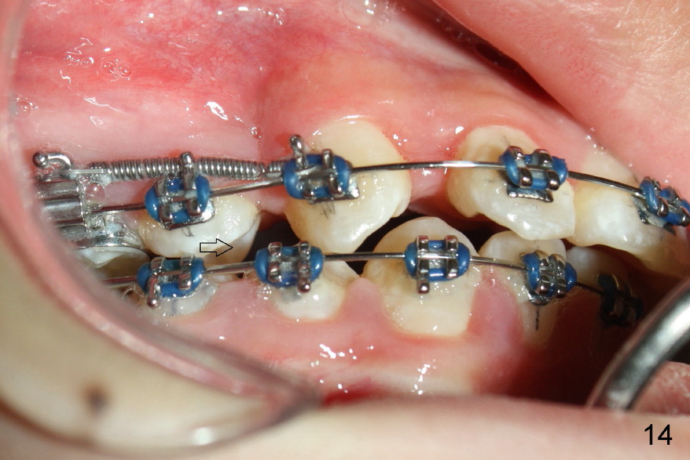
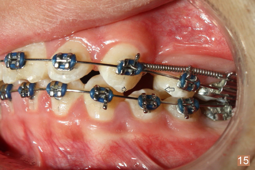
One and a half months after placement of .016x.016 niti wires and closed springs between U3 and 6, UR3 appears to have been over distalized (Fig.14), whereas UL3 needs to be further distalized. Our next main job is to mesialize U5s (Fig.14,15: large open arrows).
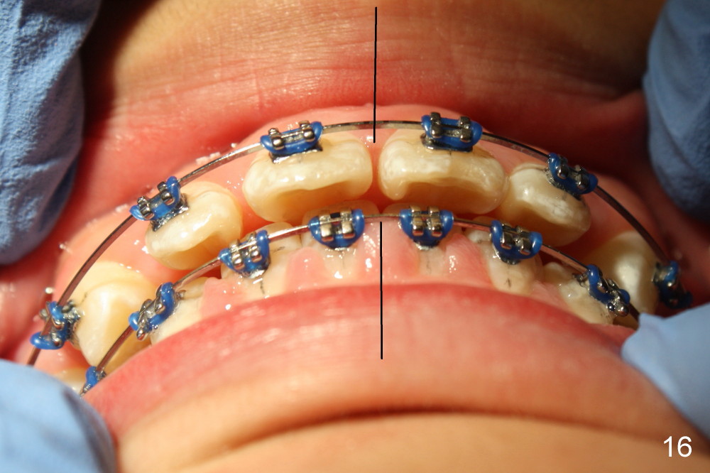
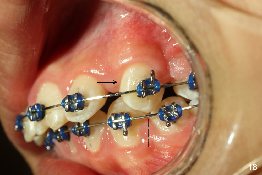
Another month of closed spring, the dental midlines nearly coincide (Fig.16) with diastemata among the upper anterior teeth. U3s move to Class I occlusion (Fig.17,18 dashed lines). .016x.022 stainless wires are placed. Next visit, a posted wire (.018x.025) is placed for the upper arch to retract the upper incisors posteriorly (Fig.17,18 arrows), followed by moving the posterior teeth anteriorly (Fig.14,15 arrows). One-year follow up is shown as follows.
Return to Ortho Cases
Xin Wei, DDS, PhD, MS 1st edition 11/23/2013, last revision 06/18/2016