|
|
|
|
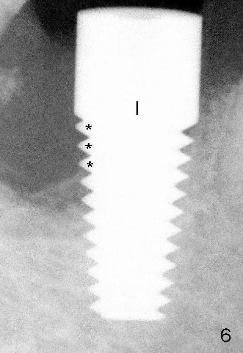
Fig.6: immediately after placement of 7x17 mm implant (I). It appears that no bone contacts the 1st three threads distally (*).
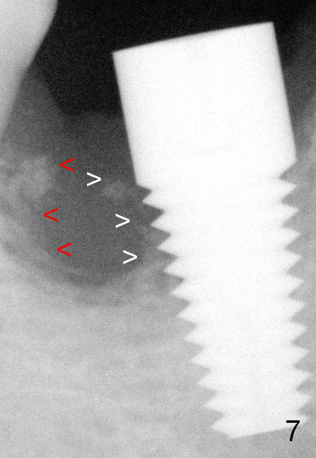
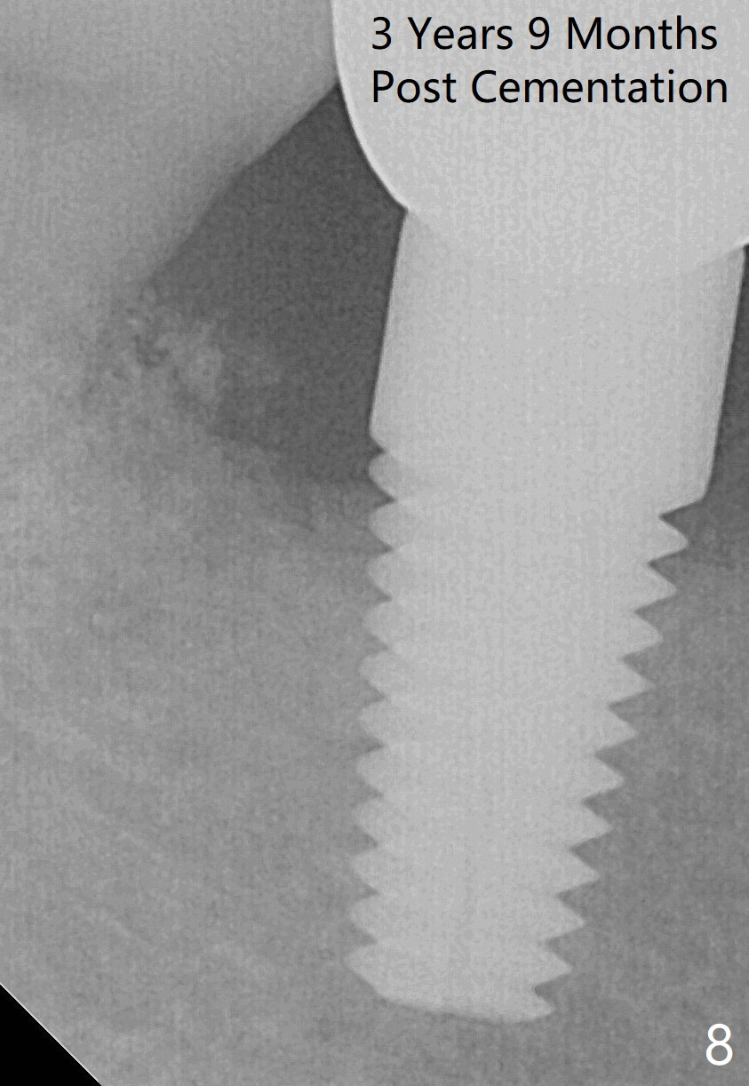
It does not look the same way 3 months 3 weeks postop (Fig.7 white arrowheads). This group of bone fragment may meet the bone indicated by red arrowheads to form the distal socket in the near future. The mesial defect appears to have been repaired 3 years 9 months post cementation (Fig.8).
Return to Septum Displacement and Regrowth Xin Wei, DDS, PhD, MS 1st edition 10/13/2013, last revision 01/28/2018