|
|
|
|
|
|
|
|
|
|
||
|
|
|
|
||
|
|
|
|
|
|
|
|
||||
Immediate Provisional
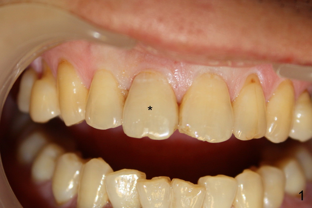
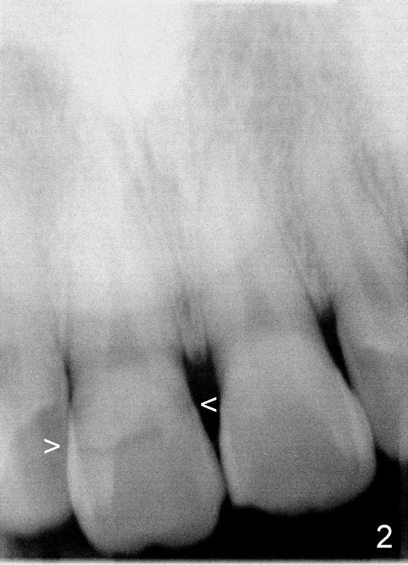
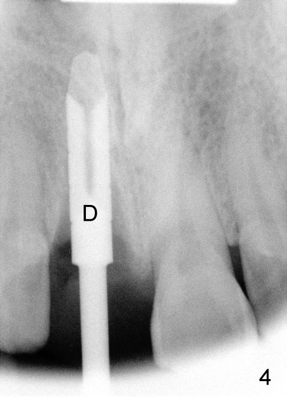
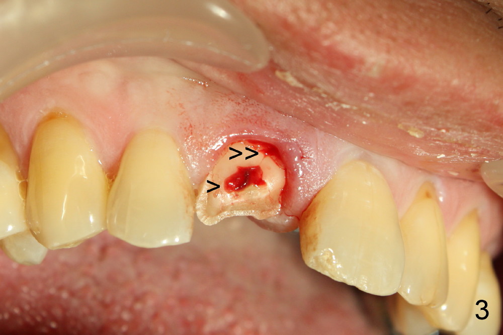
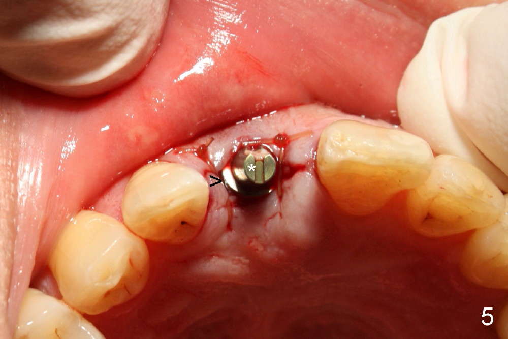
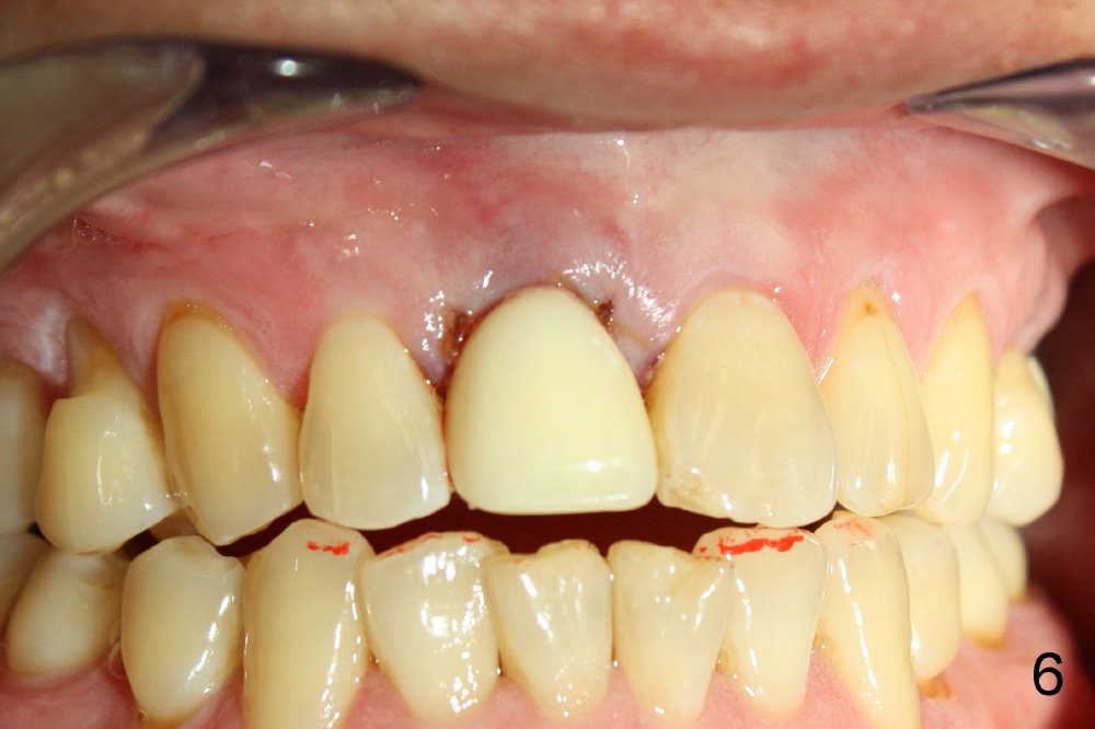
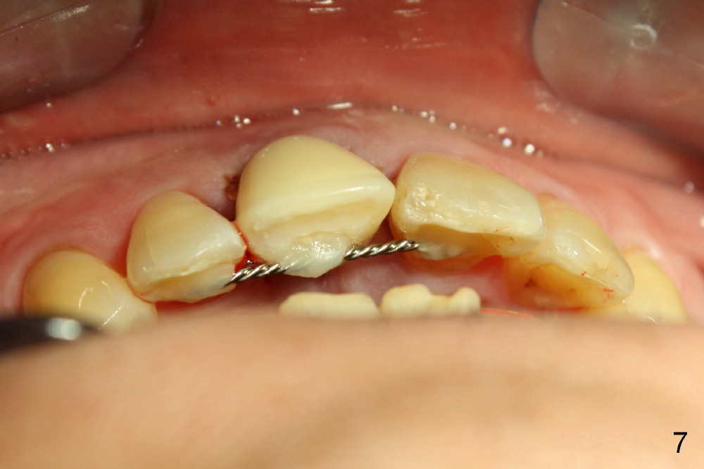
A 55-year-old man has severe pain on the upper right central incisor (Fig.1 *) after biting on a piece of bone. PA confirms crown fracture (Fig.2 <), extending subgingivally (Fig.3 >>). Osteotomy forms using a 2 mm pilot drill and 2.5-3.5 mm reamers (Fig.4). A 5x17 mm Tatum tapered implant is placed, autogenous bone placed in the buccal gap, sutures placed for wound approximation and 3.5 mm 20º angled abutment installed (Fig.5). An immediate provsional is fabricated (Fig.6), cemented and splinted (Fig.7).,
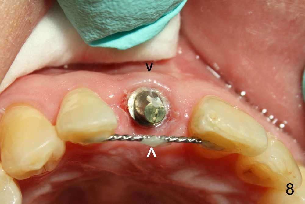
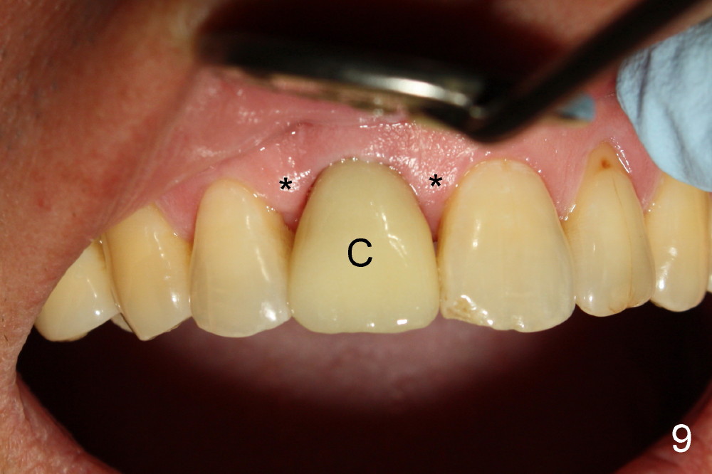
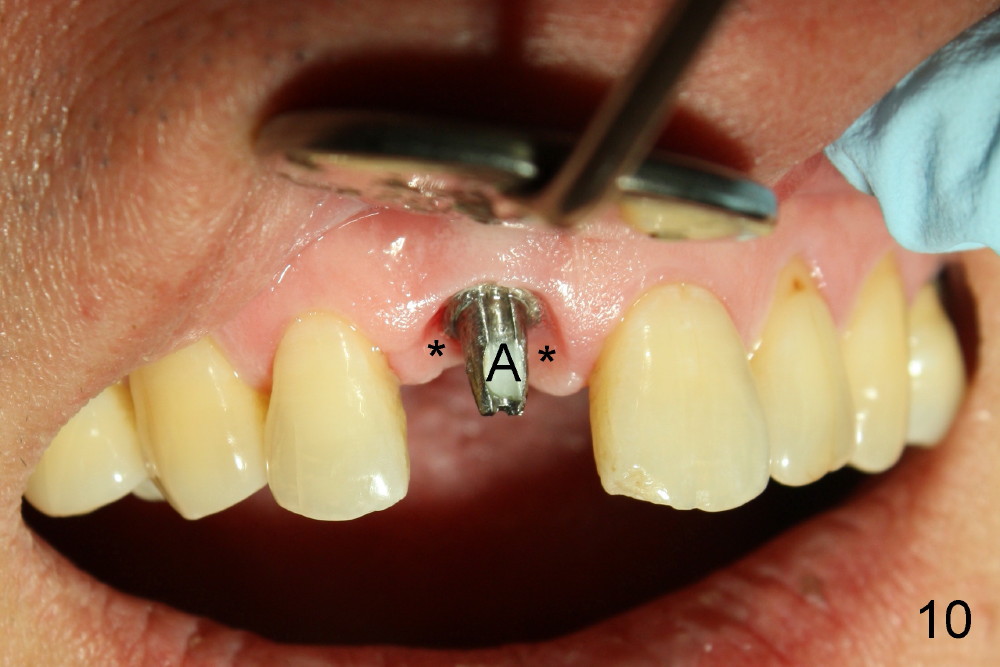
The provisional is dislodged 3.5 months postop; there is no labial atrophy (Fig.8 black arrowhead). The permanent crown (Fig.9 C) is harmonious with the papillae (*). When the try in crown is removed, the gingival tissue looks healthy (Fig.10 *).
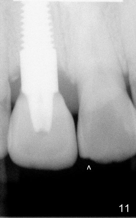
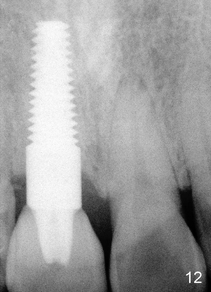
Fig.11,12 are taken 6,18 months post cementation, respectively.
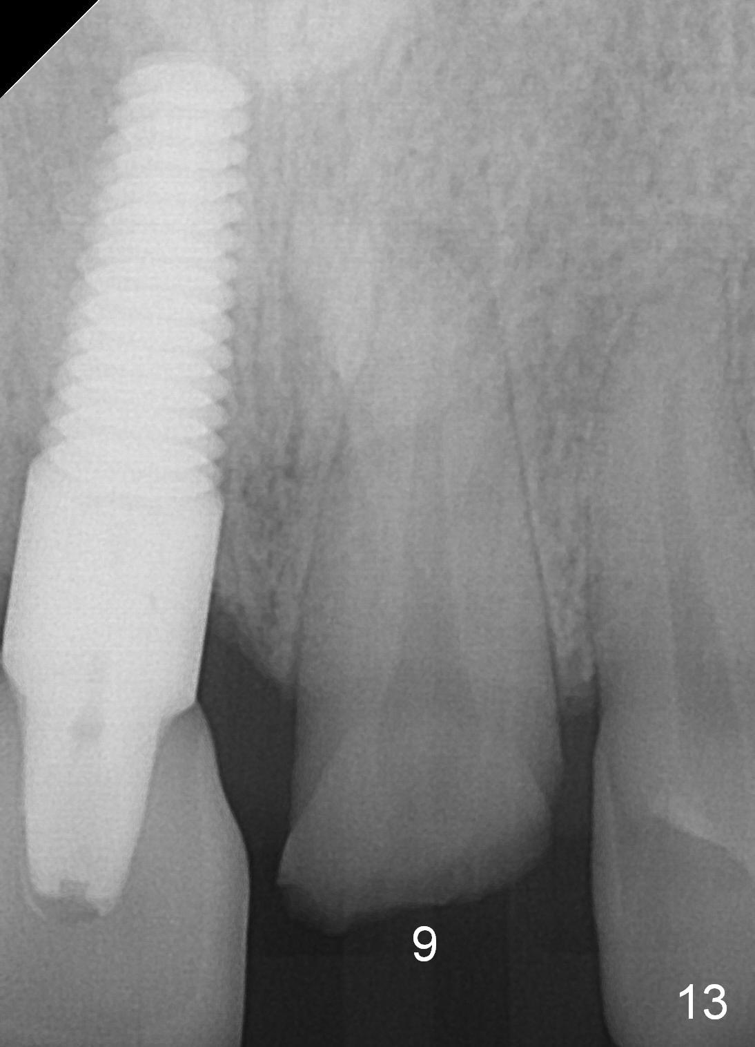
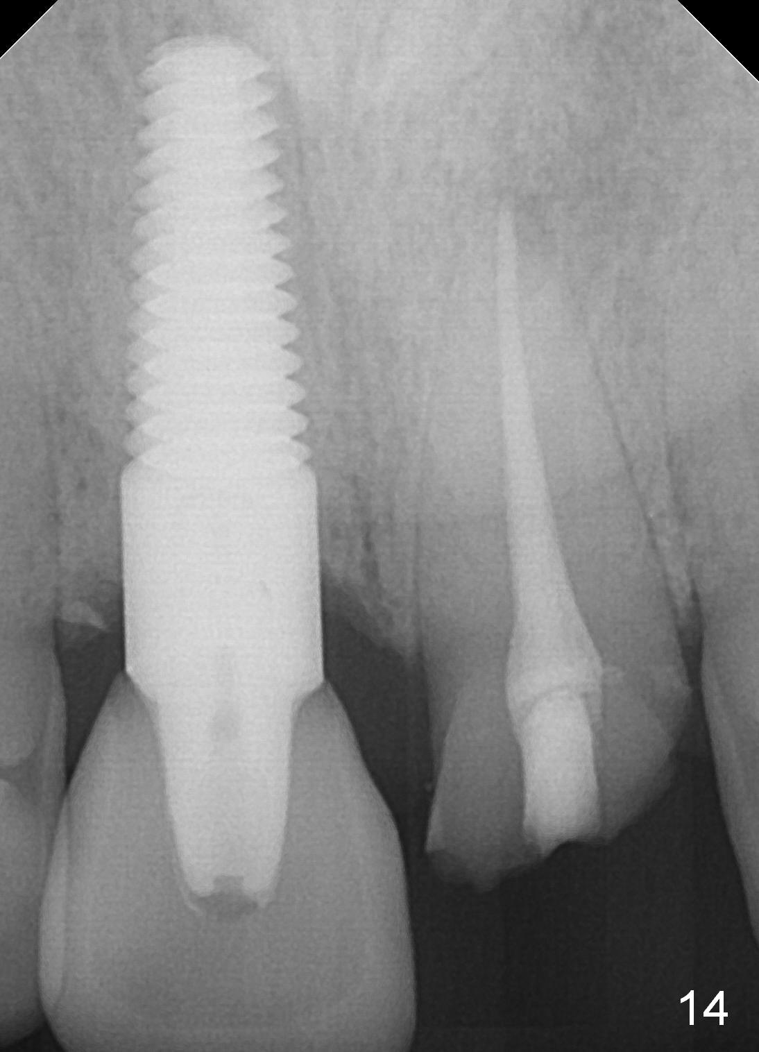
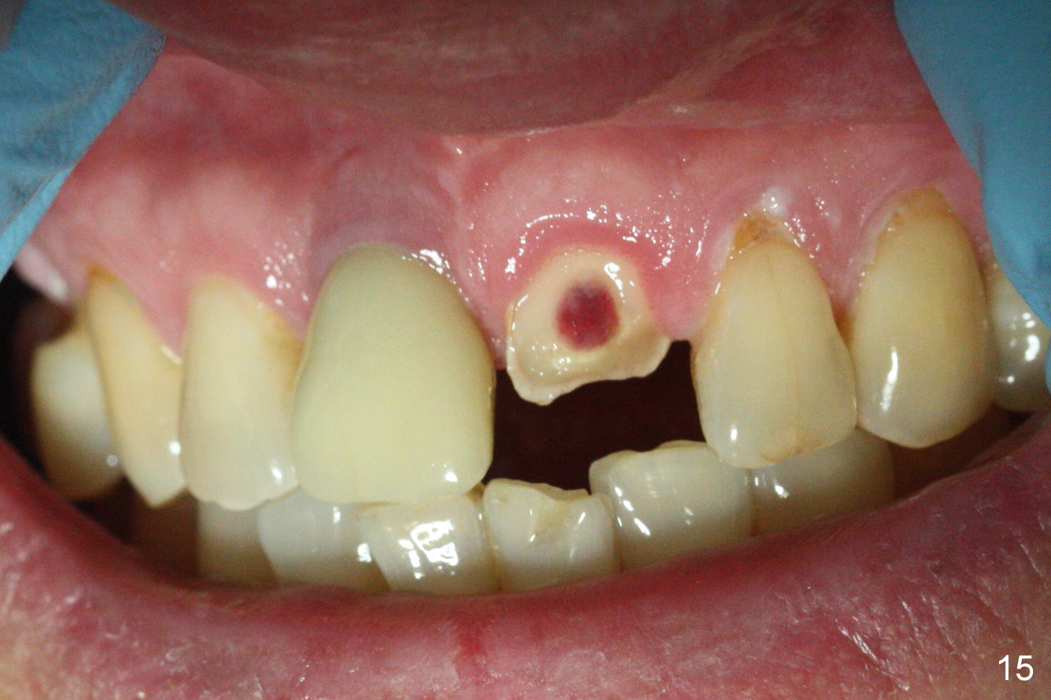
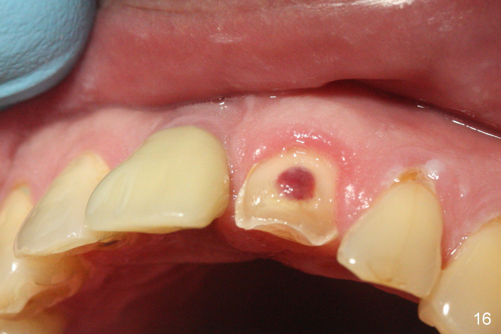
When the patient returns 3 years 10 months post cementation, the tooth #9 fractures (Fig.13,15,16), and receives root canal therapy (Fig.14). The dark gingiva could be avoided if the implant is placed more palatally and smaller in diameter (Fig.15,16).
Return to Fellowship Candidate Case Documentation Form, Upper Incisor Immediate Implant Advantages of Immediate Implant
Xin Wei, DDS, PhD, MS 1st edition 01/01/2013, last revision 07/01/2018