|
|
|
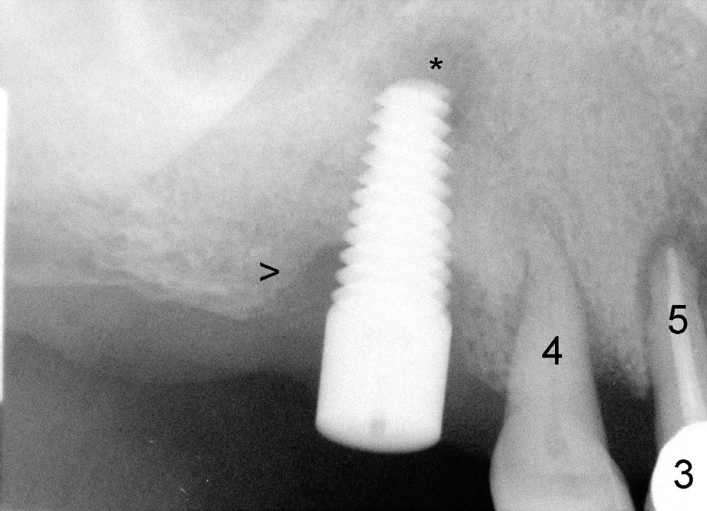
Fig.3: Four months post implantation at the site of #3. Although there is apparently bone loss at crest (>) and apex (*), the implant is stable.
The periodontal ligament space is increased for the tooth #4.
The tooth #5 has had root canal therapy and crown. There is periapical radiolucency.
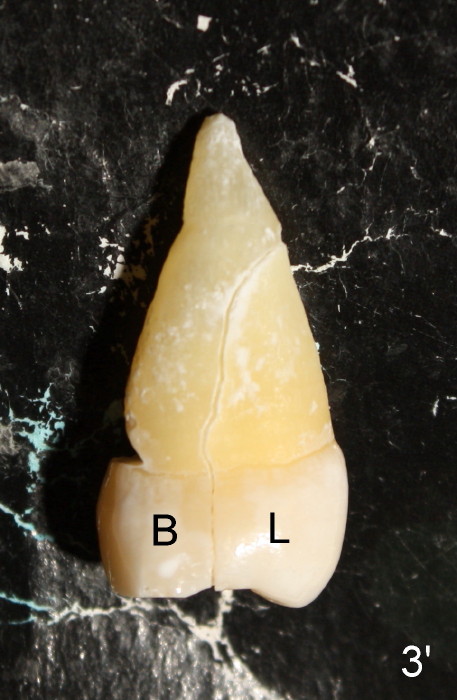
Fig.3': Extracted tooth #4 with crack line between the buccal (B) and lingual (L) portions (mesial view).
Return to Close Call
Xin Wei, DDS, PhD, MS 1st edition 06/01/2013, last revision 10/31/2014