


|
|
|
|
|
|
|
|
|
|
||
Dry Socket Prevention
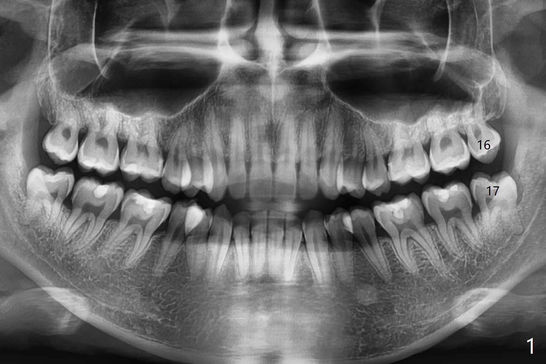
A 26-year-old man presents to clinic with pericoronitis at #17; the tooth #16 bites on the distal swollen gingiva of the tooth #17 (Fig.1). Since there appears to be no bone loss distal of #18, collagen plug, instead of Osteogen plug (Fig.2,3), will be placed in the socket(s) after extraction (Fig.4,5). For easy insertion, the plug is cut apically (Fig.6 *). The wound is closed with 4-0 plain gut suture. The patient returns for #1 and 32 extraction, eight months post #16 and 17 extraction. As usual, no bone substitute is placed in #1 socket after extraction (Fig.7). After #32 extraction, the distal socket looks large so that Augma (Bond Apatite) is placed and pressed (A), followed by a piece of Collagen Plug (C, to prevent Augma dissolved by saliva in case premature loss of suture). In fact there is also buccal defect at #32 due to chronic infection and heavy calculus. There is cortical bone formation at #16 socket opening (^). The mesial and distal sockets of #17 (*) seem to be obliterated because of placement of Collagen Plug. Return to Plug, Weichat Xin Wei, DDS, PhD, MS 1st edition 01/05/2019, last revision 09/22/2019