|
|
|
|
|
|
|
|
|
|
Cocktail Graft
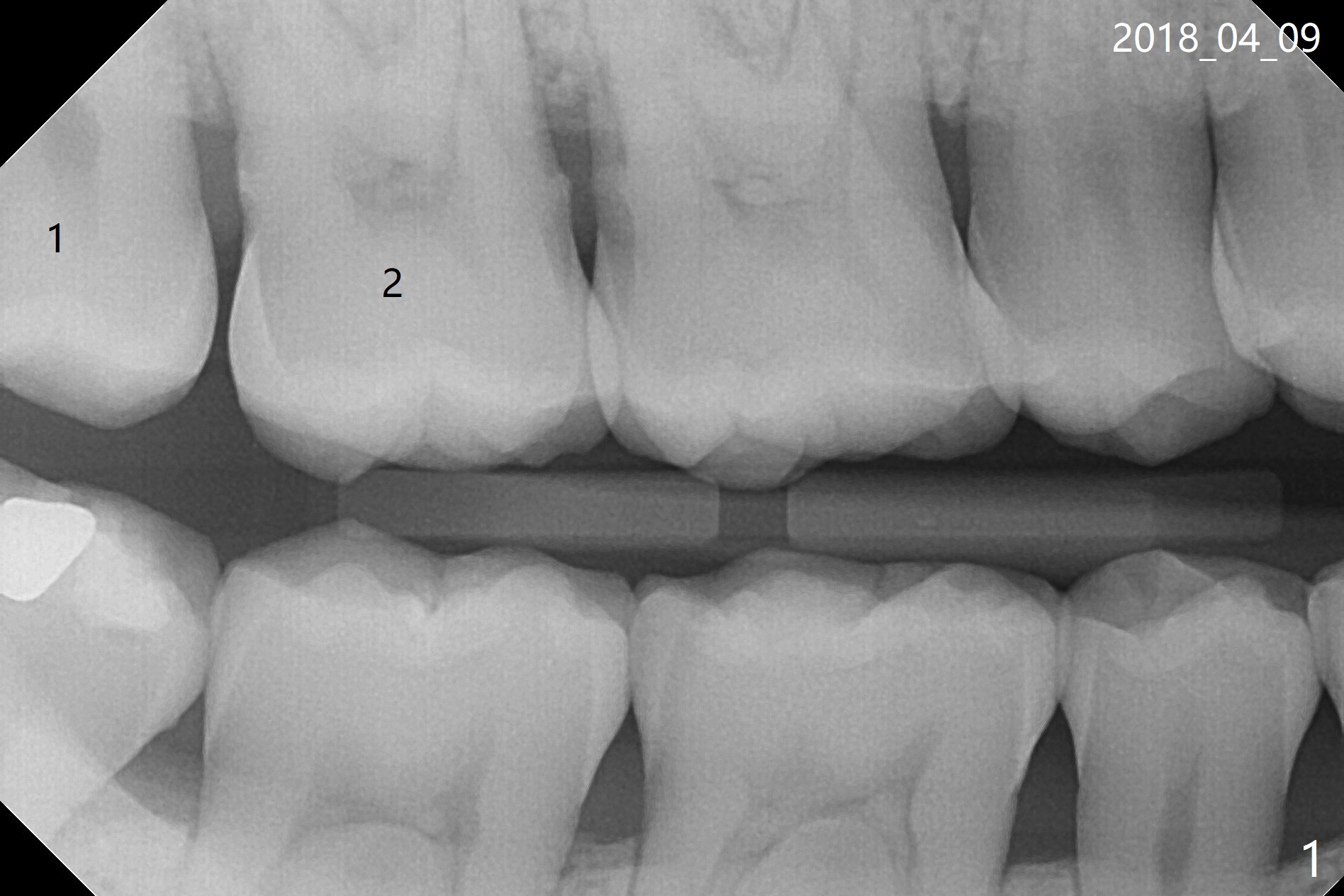
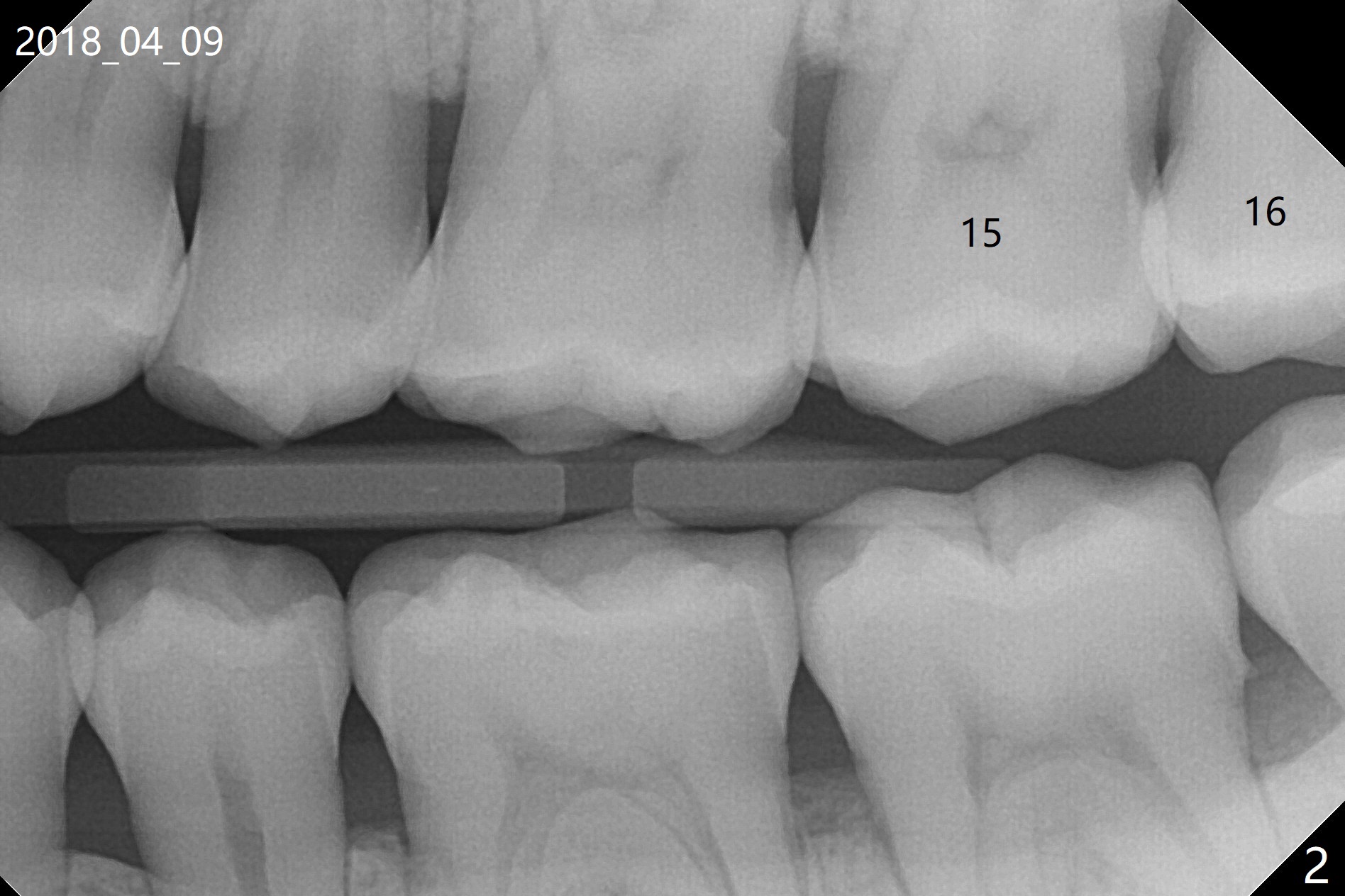
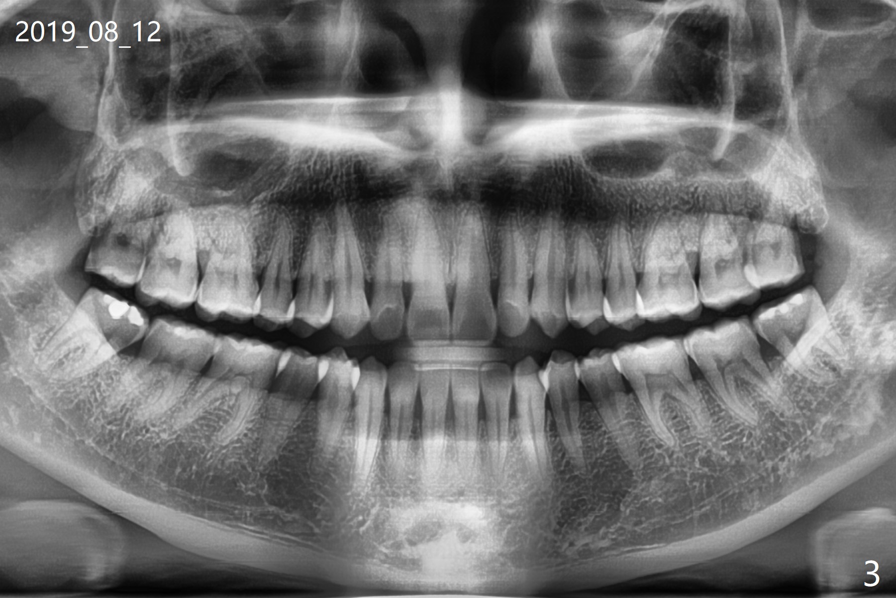
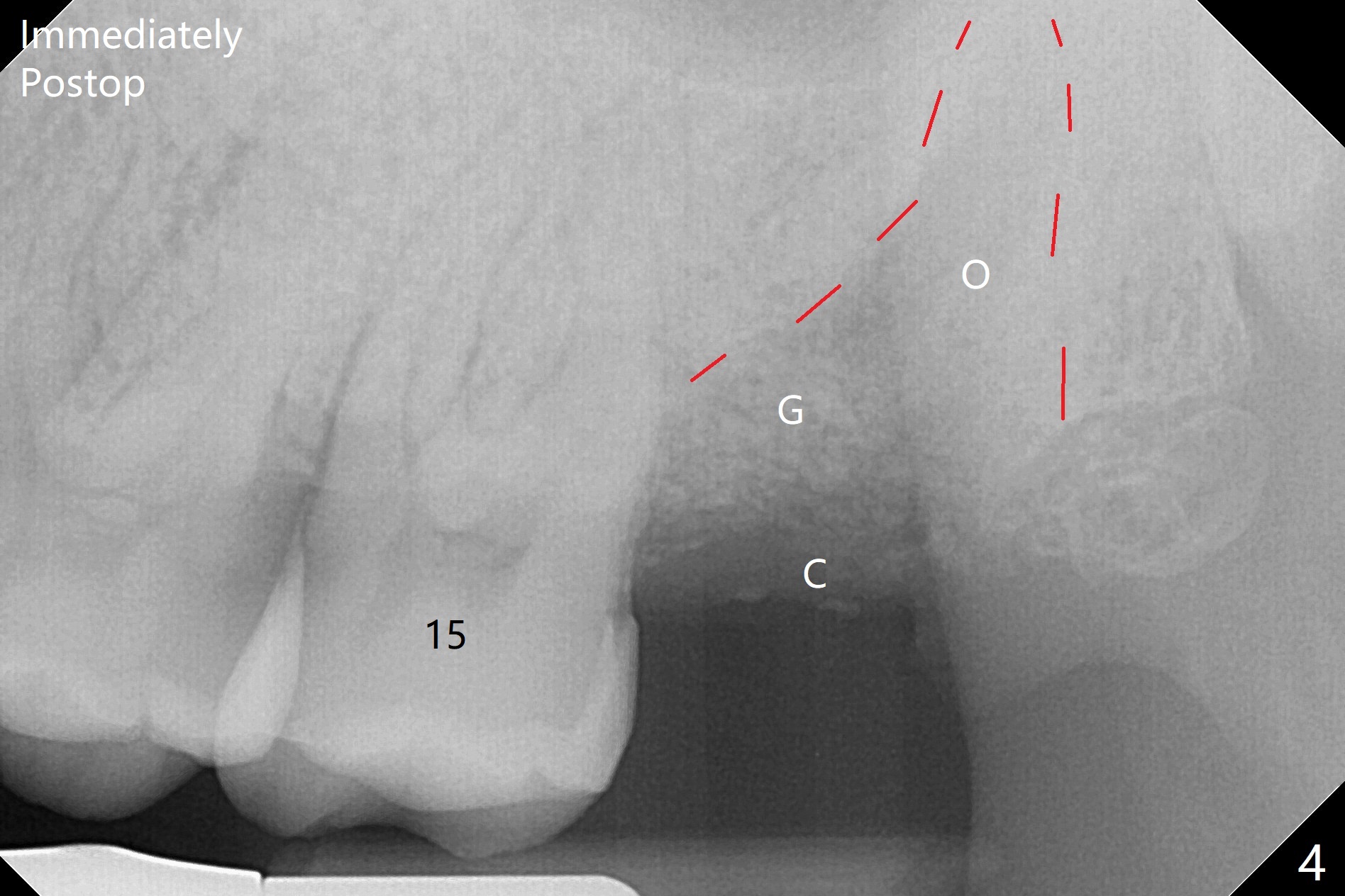
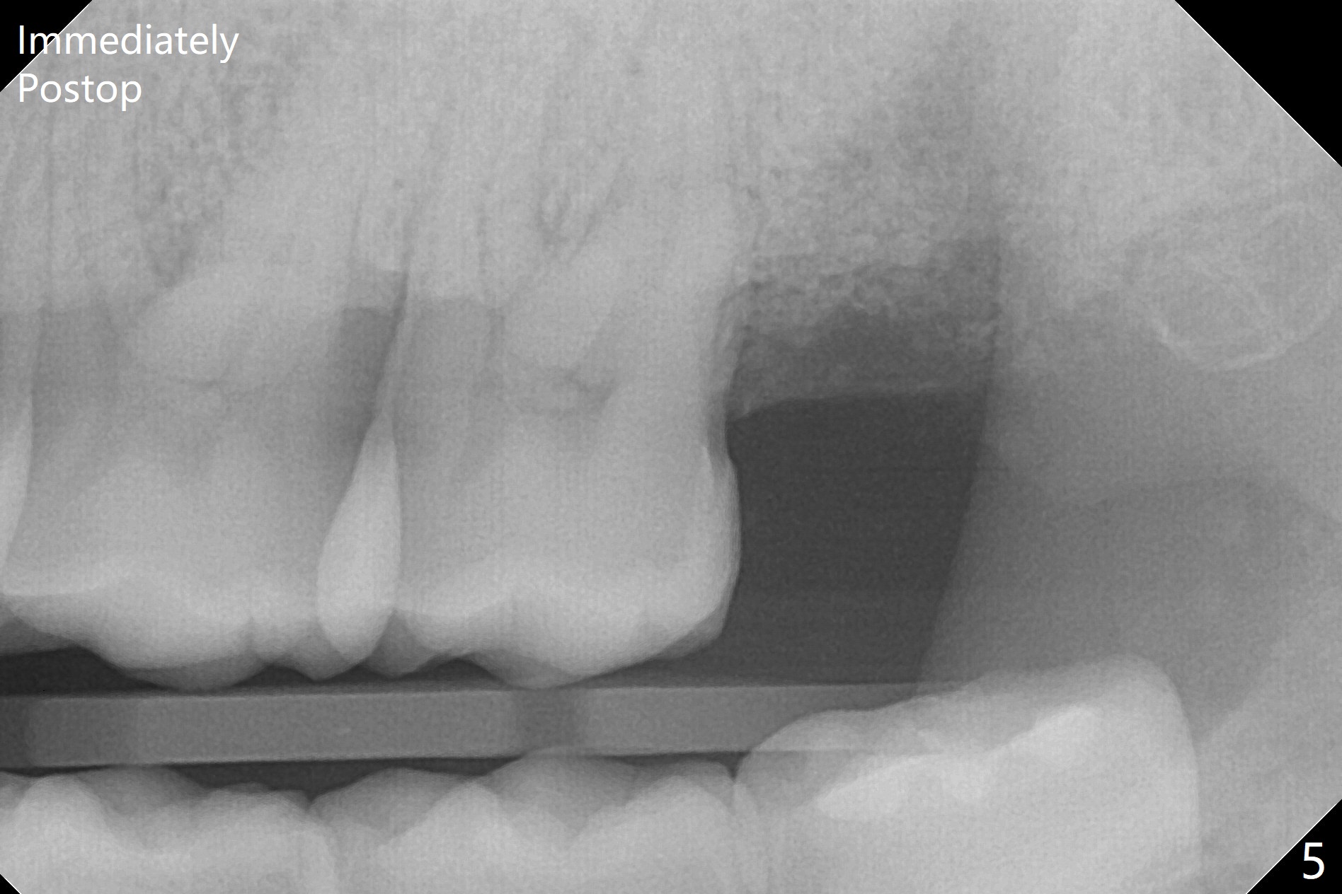
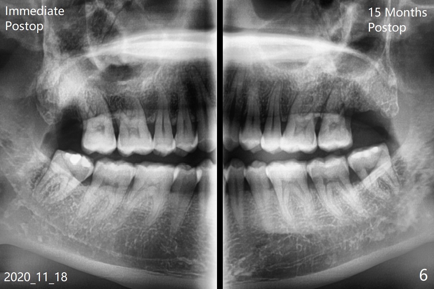
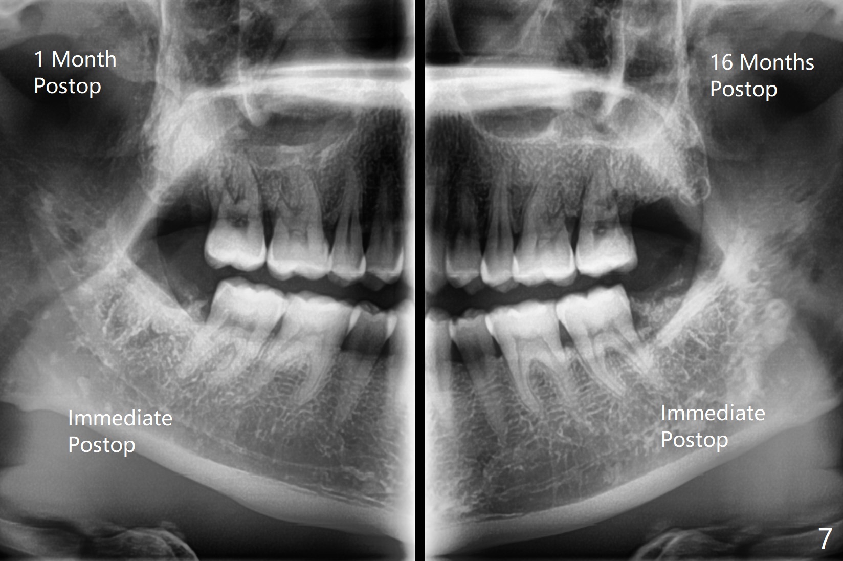
A 35-year-old woman requests extraction of the tooth #1 (food impaction between #1 and 2, Fig.1) and 16 (pain and 6 mm pockets between #15 and 16, Fig.2). In spite of the fact that there is bone loss between #14 and 15 (Fig.3), there is no deep pocket between them. After extraction of #16, SRP is performed in the distal surface of #15 with removal of granulation tissue and application of Endogain. Osteogen plug (Fig.4 O) is placed in the apical and distal portion of the socket of #16 (red dashed line), while allograft (G) against the distal root surface of #15. Finally Collagen plug (C) and 6-month membrane are placed in the opening of the socket, followed by suturing and periodontal dressing. The bone density is high post #1 extraction (Fig.6), related to difficult removal. It appears that bone graft at #16 (^) remains in place 15 months postop. Since no buccal trough is made for extraction, the external oblique ridge is present at #17 and 32 before and after surgery. Bond Apatite is placed with Collagen Plug (Fig.7).
Return to
Plug
Xin Wei, DDS, PhD, MS 1st edition
08/29/2019, last revision
12/19/2020