|
|
|
|
|
|
|
|
Bone Graft for Periimplantitis While 3rd Molar Extraction M
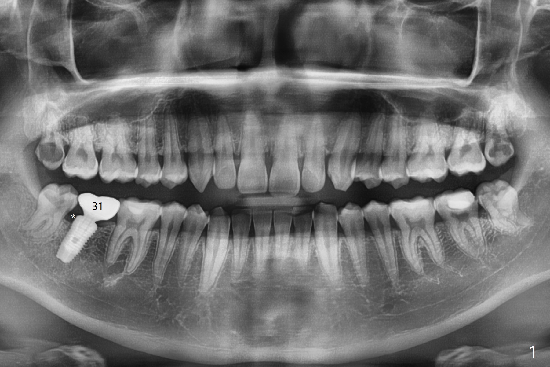
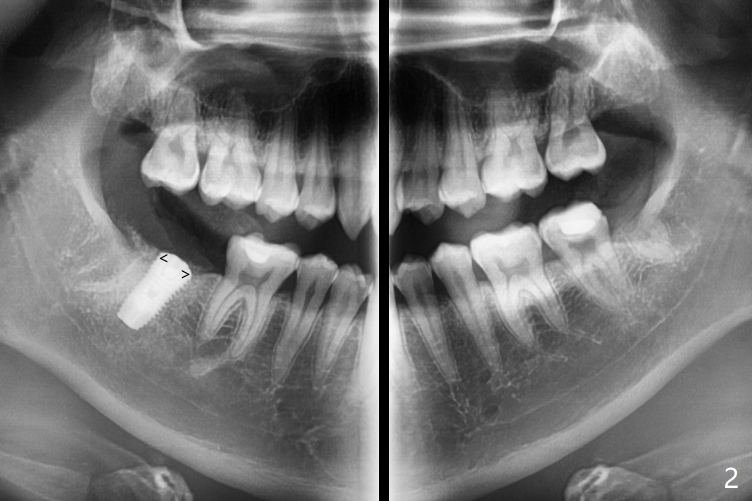
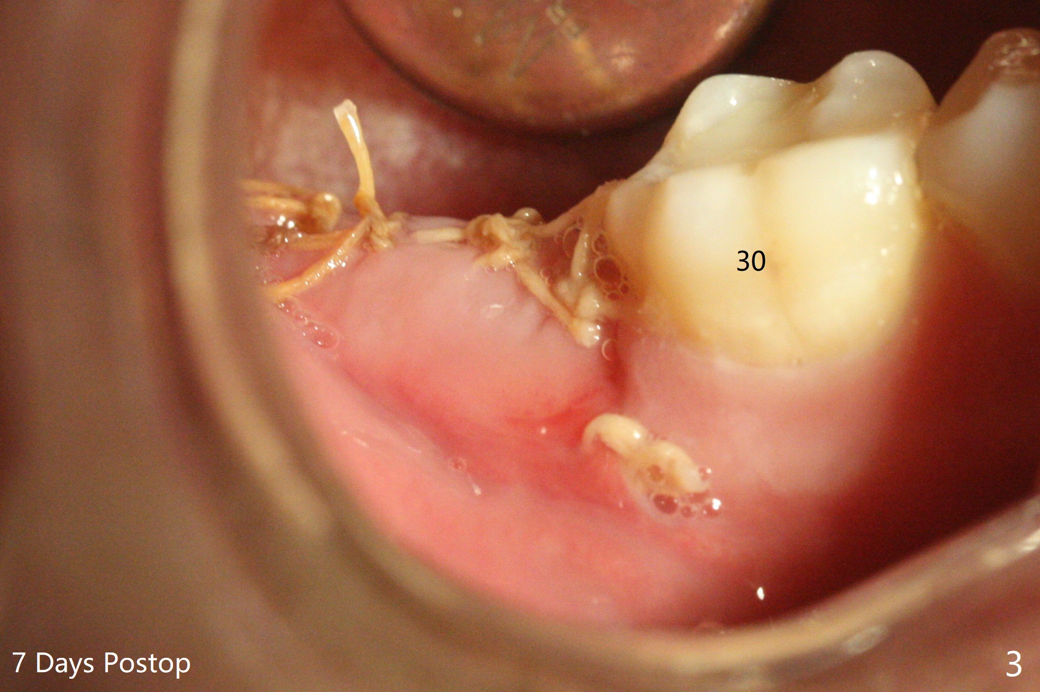
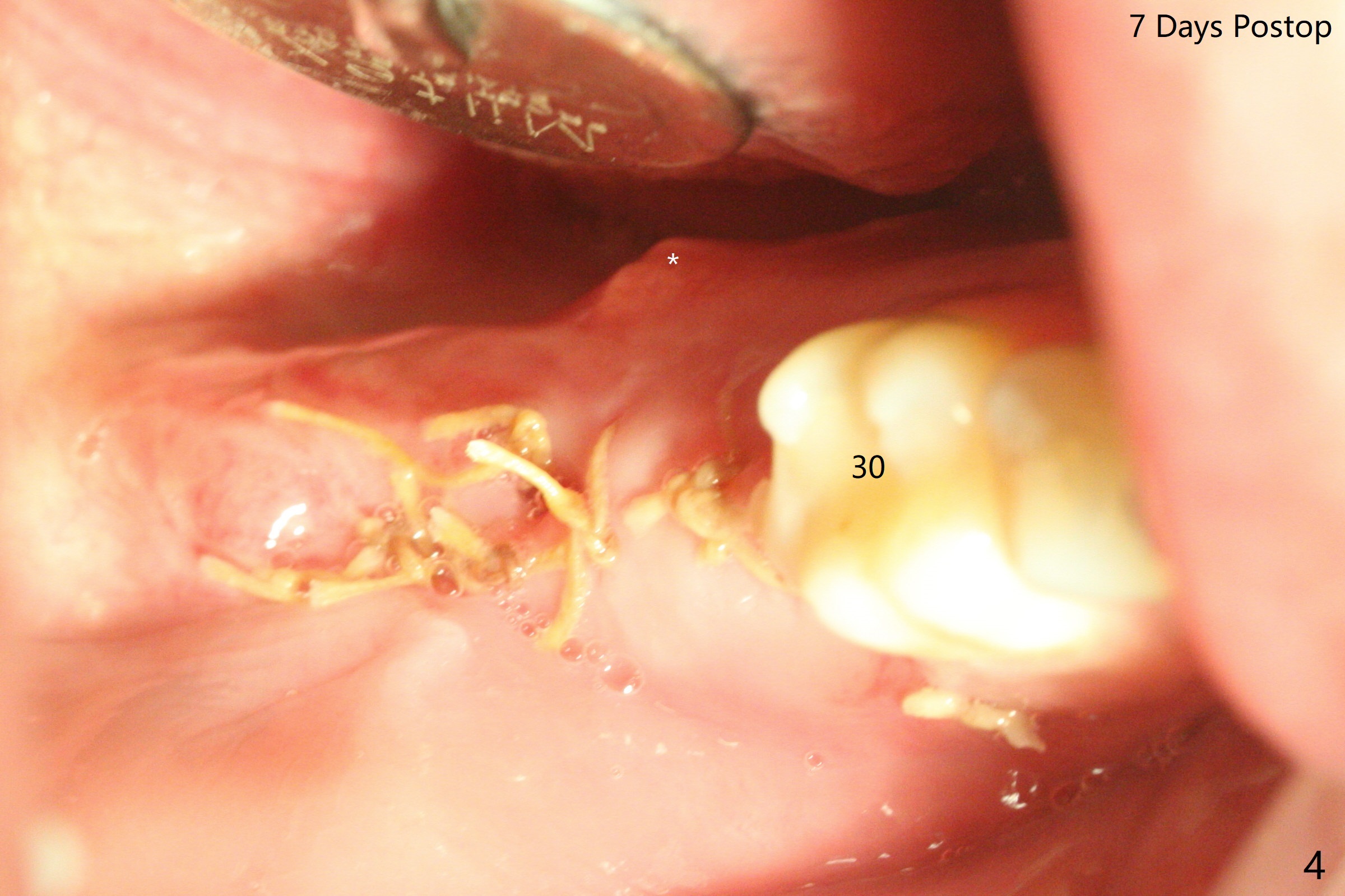
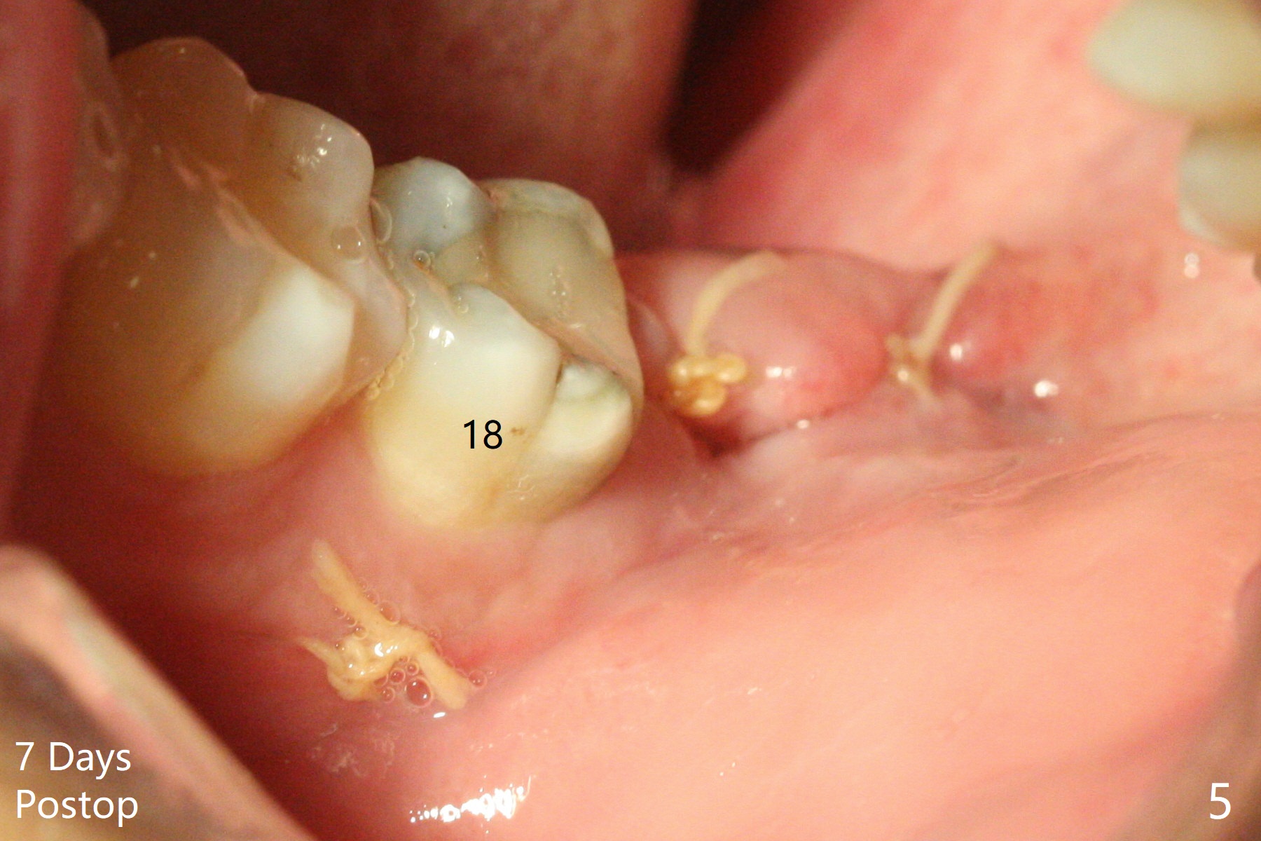
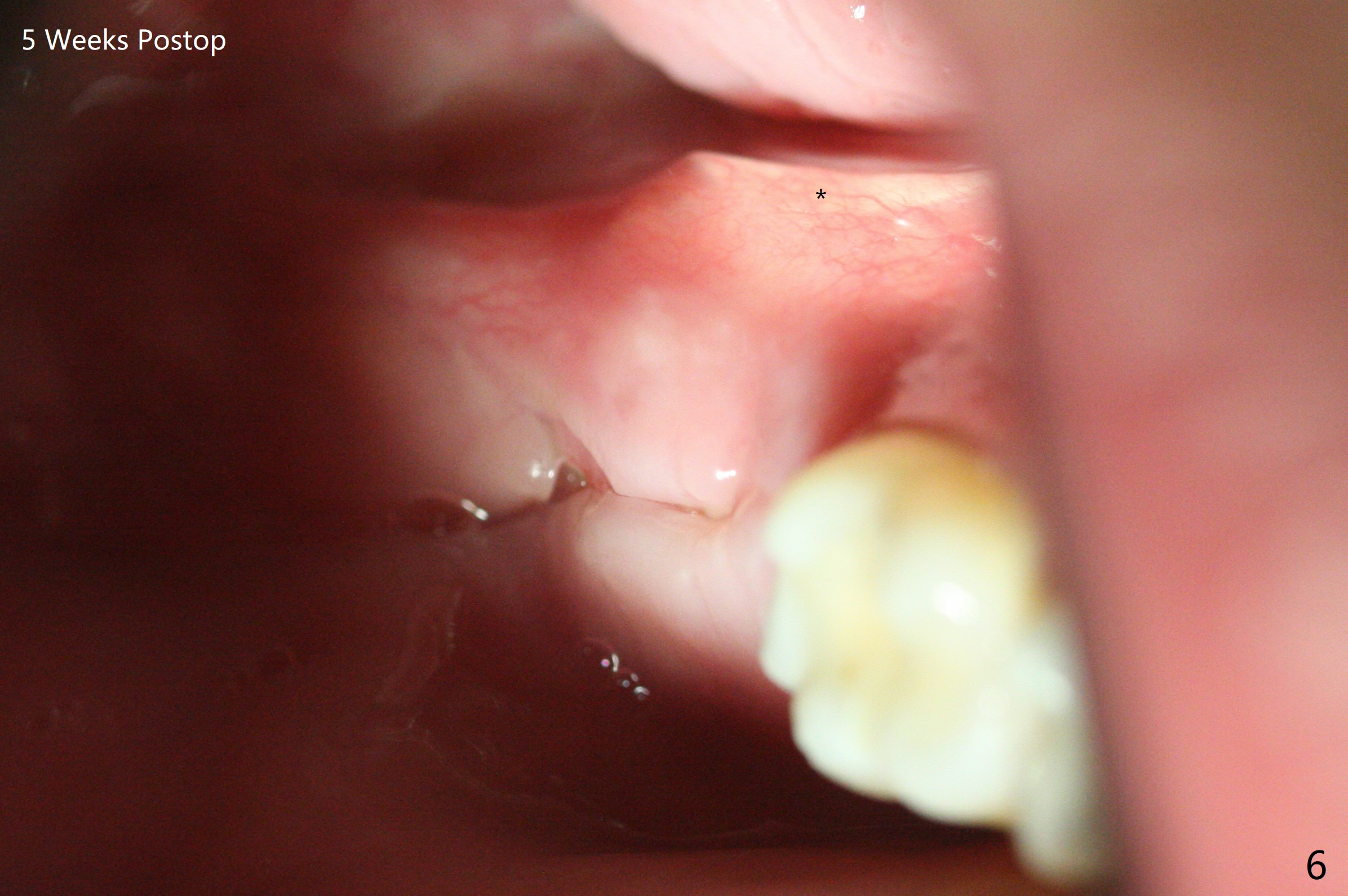
A 26-year-old man returns for #17,32 extraction 5.5 months post #1,16 one (Fig.1,2). In fact the crown/abutment at #31 is loose and removed, which makes easy for access, debridement with Titanium brush and bone graft (Fig.2 arrowheads, Ossogen). Osteogen plug is placed in the extraction sockets. The patient has limited mouth opening. Hemorrhage in #17 socket is controlled apparently with the plug following incomplete removal of granulation tissue. A piece of 12x12 mm Amnion-Chorion Membrane seems small for bone graft coverage. Cytoplast is added, followed by PGA suture. The former may keep the bone graft in place in case the wound dehisces. In fact the implant was not placed deep enough. It should have been removed. Although the wounds at #31/32 and 17 heal 7 days postop (Fig.3,5), the Cytoplast is visible immediately underneath the lingual gingiva at #31 (Fig.4 * (bulging)). The patient returns for Cytoplast (Fig.6 (occlusal view) *) removal 5 weeks postop. The procedure is done smoothly.
Return to Plug Allograft Xin Wei, DDS, PhD, MS 1st edition 12/24/2019, last revision 02/01/2020