|
|
|
|
|
|
|
|
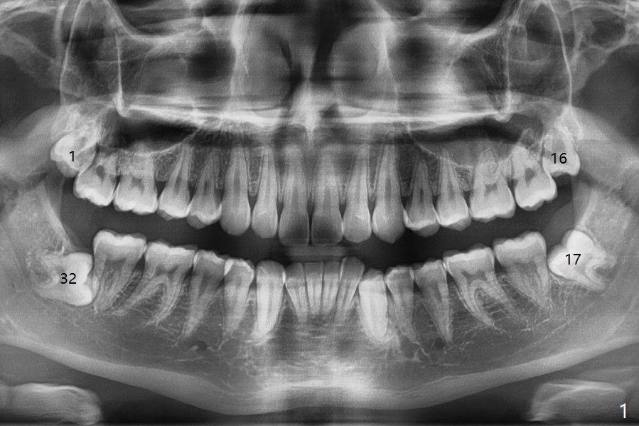
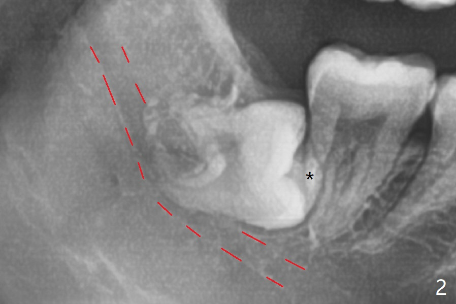
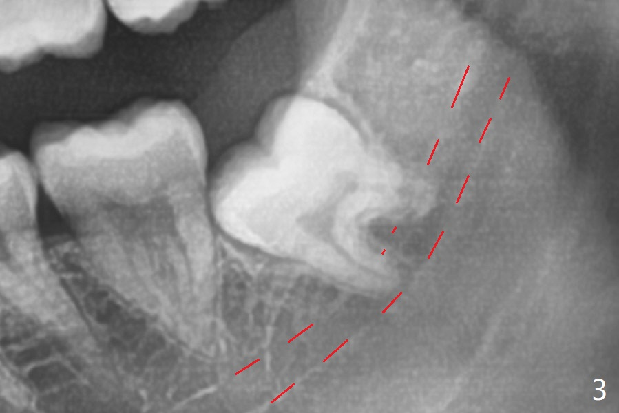
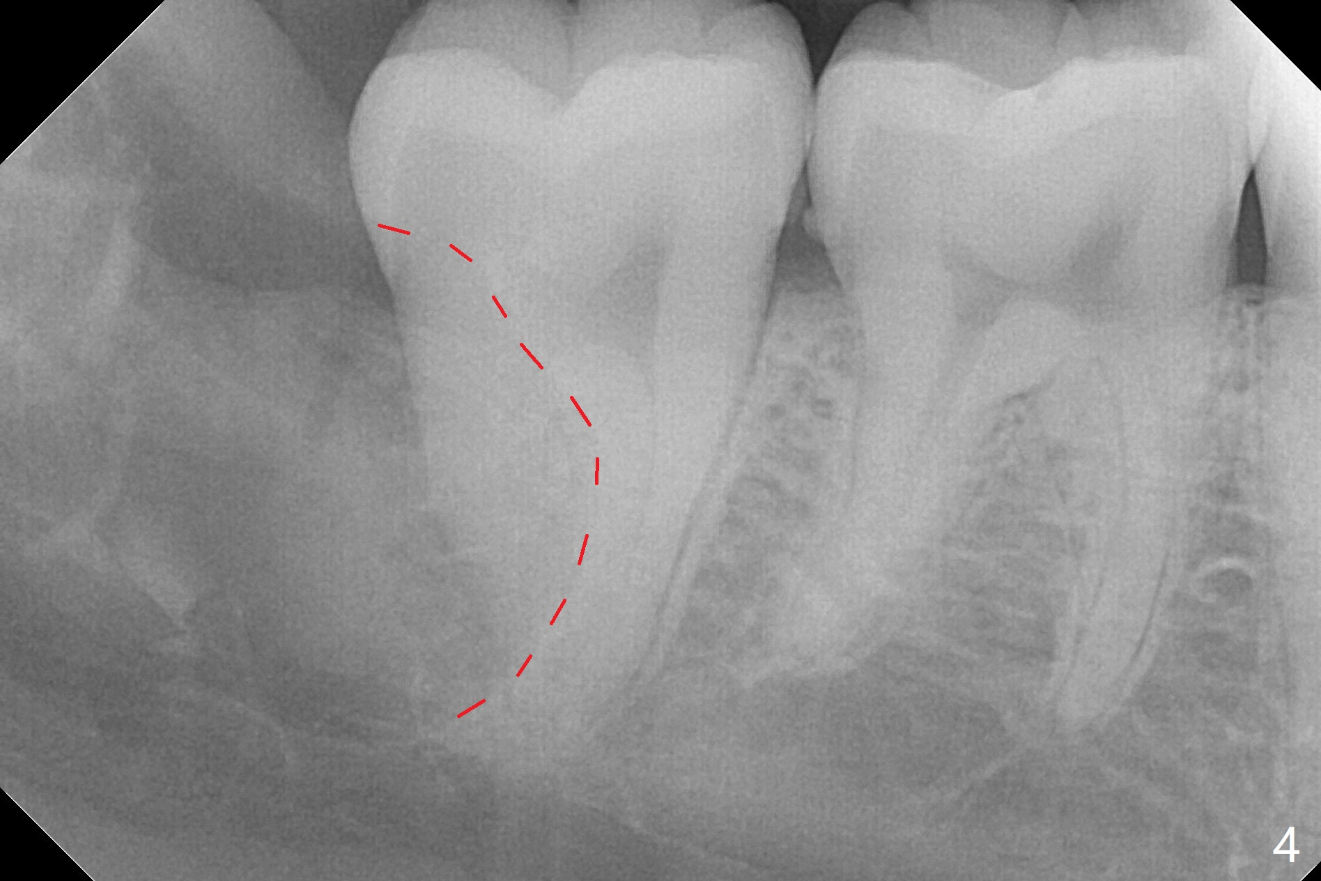
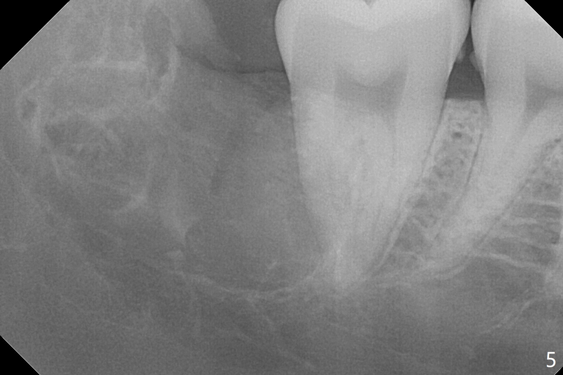
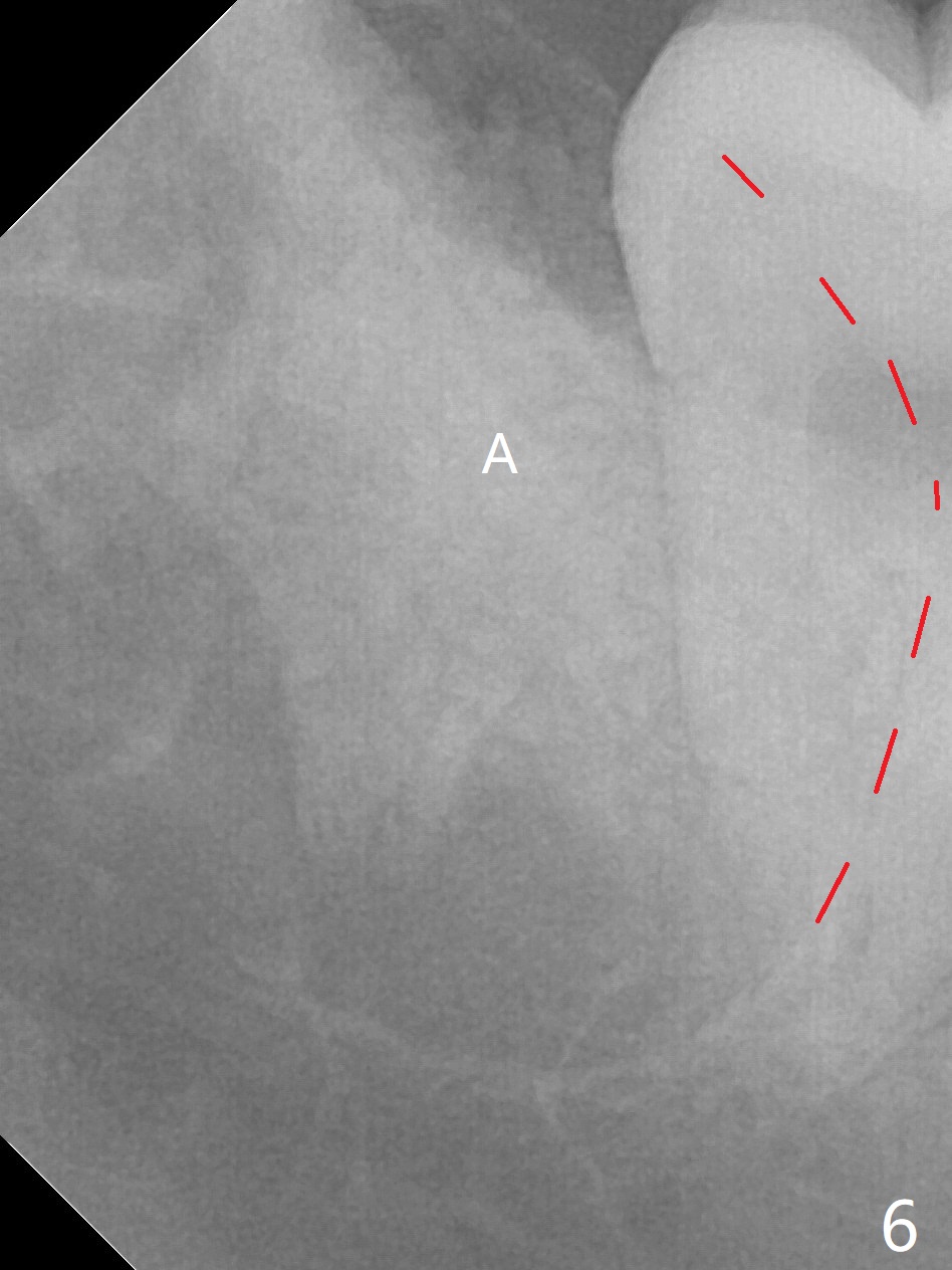
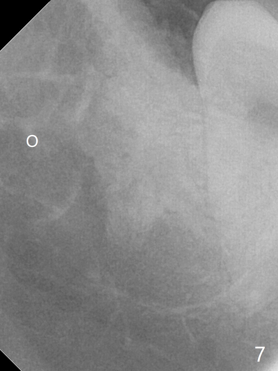
Half of Augma in Distal Sockets of Lower 3rd Molars
A 17-year-old man will return for extraction of 4 of the 3rd molars (Fig.1). Since the distobuccal surface of the tooth #31 may lack the bone (Fig.2 *), half of Augma will be placed in the distal portion of the sockets of #17 and 32, while allograft in the mesial one. Take PAs immediately post extraction to determine bony defects of the lower 3rd molars. Insert a piece of 2x2 gauze into the socket immediately post extraction. If hemorrhage is severe because of closeness between the mesial roots of the lower 3rd molars and the Inferior Alveolar Canal (Fig.2,3 red dashed line) after removal of the gauze, insert a half piece of Collagen Plug or more into the socket. If the latter is effective in hemostasis, Augma will be not applied, but allograft will be used, followed by Collagen Plug on the top of the allograft and sutures. Prepare both 4-0 and 5-0 ones. In fact the patient chooses to have 2 of the 3rd molars to be extracted. The extraction at #32 turns out to be extremely difficult with numerous sectioning because the crown of #32 is embedded into the distolingual surface of the fused conical roots of #31 (Fig.4,5). After insertion of a whole piece of Osteogen plug (Fig.7 O) into the root portion of the socket, Bond Apatite (1 cc) is pressed into the coronal portion of the socket (Fig.6 A). Due to oozing, the cement does not seem to be set completely. One third piece of Collagen plug is placed on the top of the cement before 4-0 Chromic gut suturing tightly. CBCT will be taken for better treatment approach prior to #17 extraction. Return to Plug Augma Xin Wei, DDS, PhD, MS 1st edition 10/06/2019, last revision 12/28/2019