|
|
|
|
|
|
|
|
|
|
|
|
|
|
|
|
|
|
3D Bond vs. Osteogen Plug I
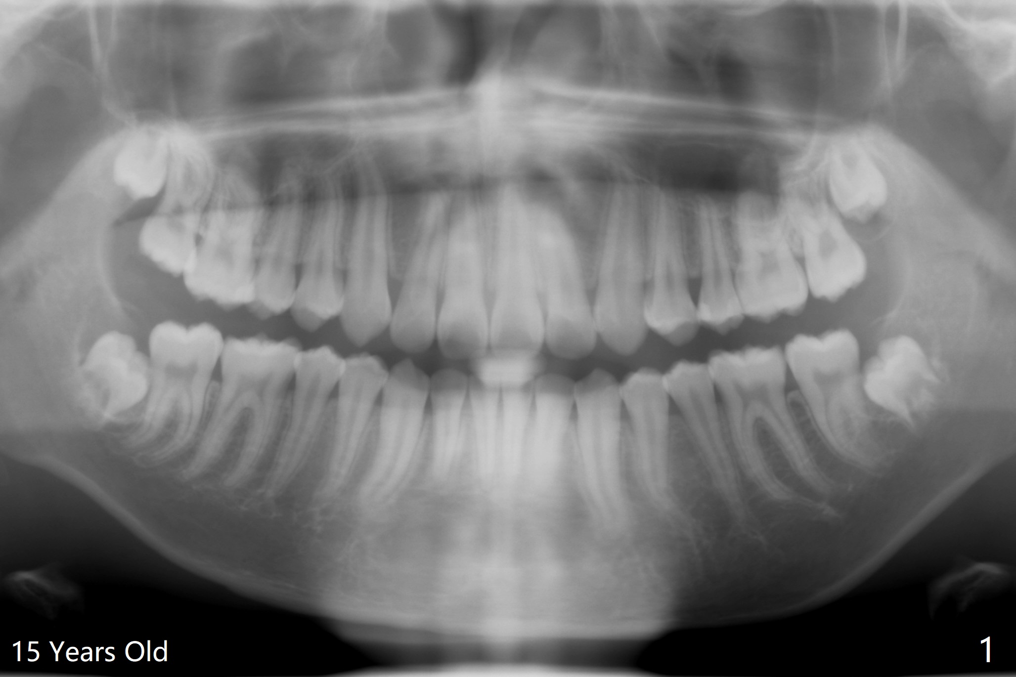
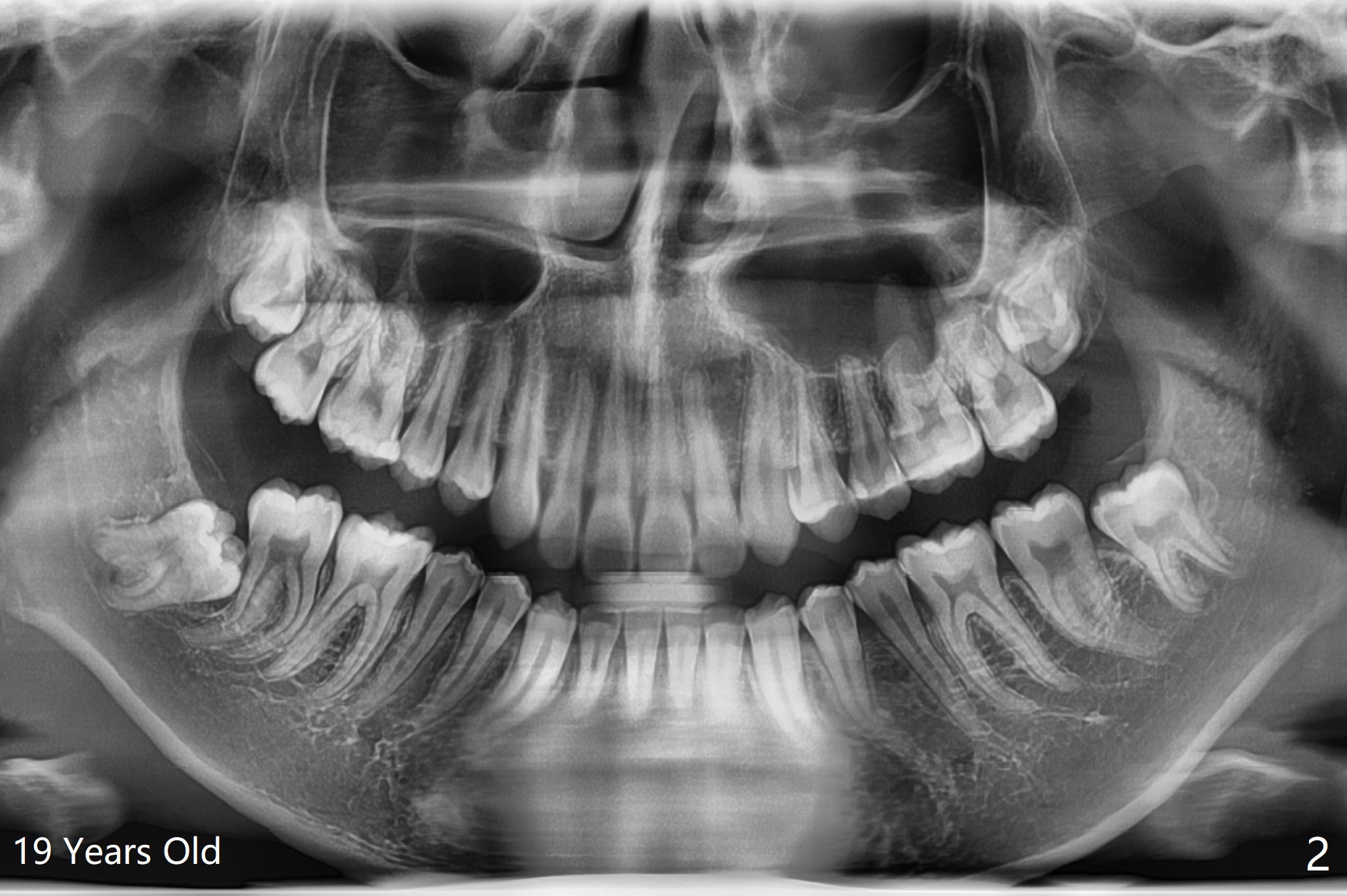
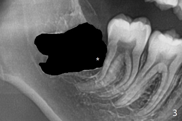
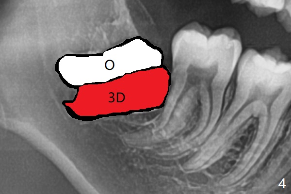
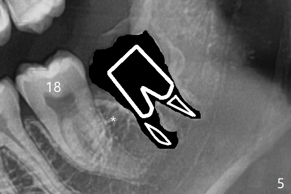
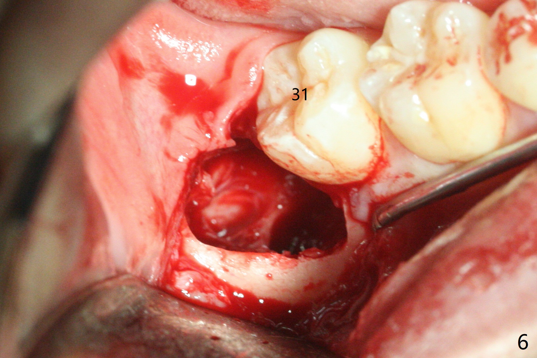
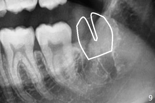
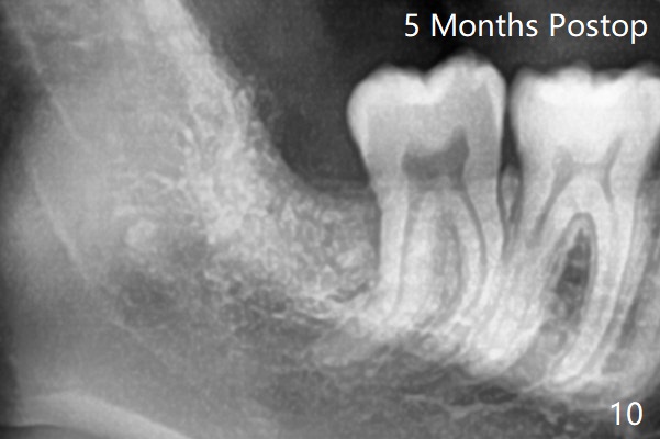
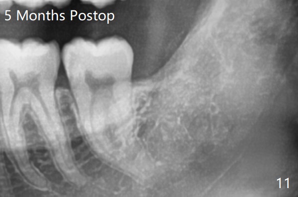
A 19-year-old man returns for #1,16,17 and 32 extraction (Fig.1,2). Because of horizontal impaction of #32 and no bone #31 distal post extraction (Fig.3 *), 3D bond (.5 cc) will be placed in the mesial socket of #32 (Fig.4 red), while Osteogen plug (1/2 (O)) in the distal one. Since there is no bony defect between #17 and 18 after extraction (Fig.5 *), a piece of Osteogen plug (cut half apical) will be inserted in #17 socket, whereas 2 small pieces of Osteogen plug placed in the apical portions (triangle and spindle shaped). Take posterior panoramic X-ray postop (bitewing type, CT format, not XV). There appears bone formation in the socket 5 months postop (Fig.10,11).
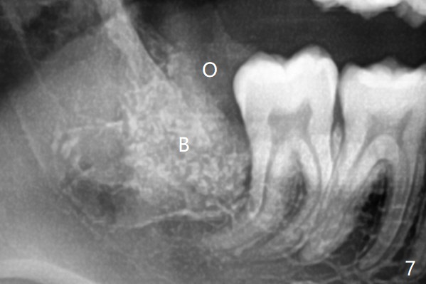
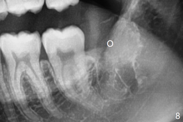
Since #32 sockets seem large, extending mesiobuccal (Fig.6), Bond Apatite (1 cc) is placed instead (Fig.7 B), covered by 1/3 of Osteogen plug (O) and sutured with 4-0 PGA. Extraction of #17 is also difficult. As 2 sockets are indistinct, a piece of Osteogen plug with split is placed (Fig.8) upside down (Fig.9 vs. Fig.5). There appears bone formation in the socket 5 months postop (Fig.10,11).
Return to
Plug
Xin Wei, DDS, PhD, MS 1st edition
03/07/2020, last revision
08/01/2020