
|
|
|
|
|
|
Allograft & Osteogen Plug
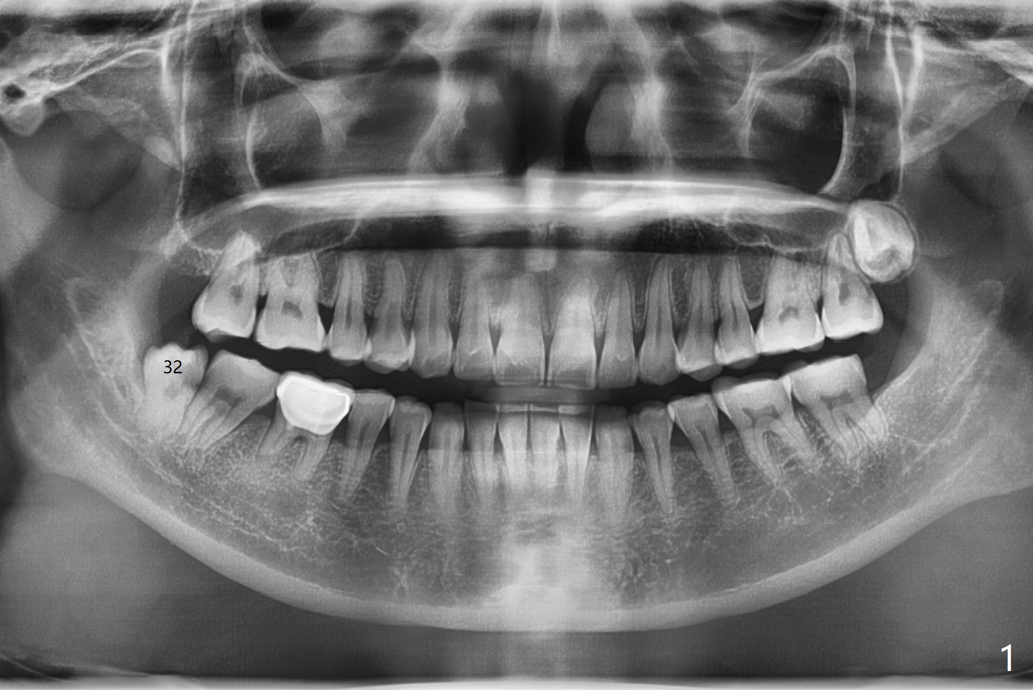
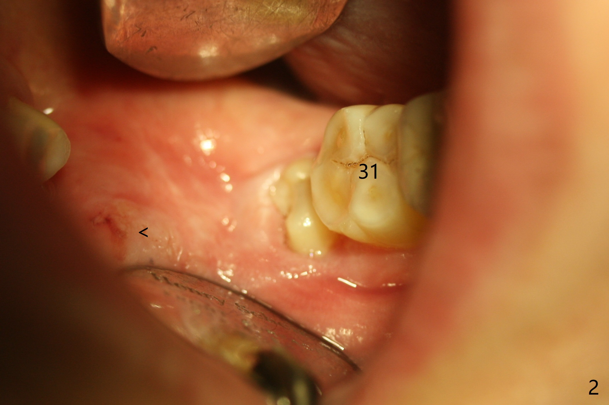
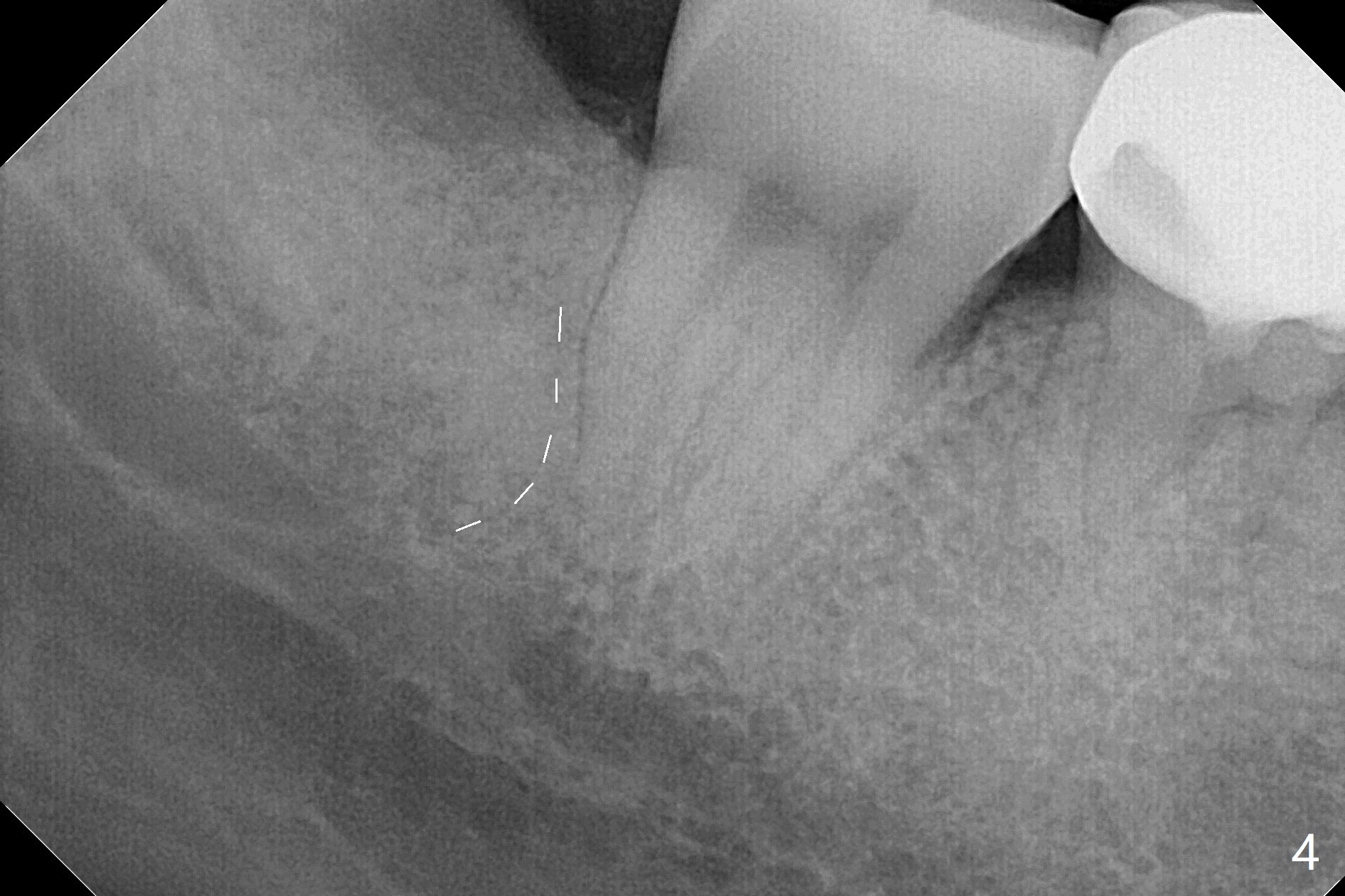
A 37-year-old woman requests extraction of the tooth #32 (Fig.1,2 (< cheek bite)). Note no bone between the last 2 molars. The tooth is extracted with incision, but without bone removal. Vanilla graft (Fig.3 *) is placed as mesial as possible, while Osteogen plug (P) coronal before suturing. The white dashed line in Fig.4 most likely represents the mesial wall of the 3rd molar socket (fused roots). The distal root of the 2nd molar seems to be partially covered by the bone.
Return to
Plug,
Weichat
2Xin Wei, DDS, PhD, MS 1st edition
01/30/2019, last revision
07/17/2021