
|
|
|
|
|
|
|
|
|
Porcelain Chips
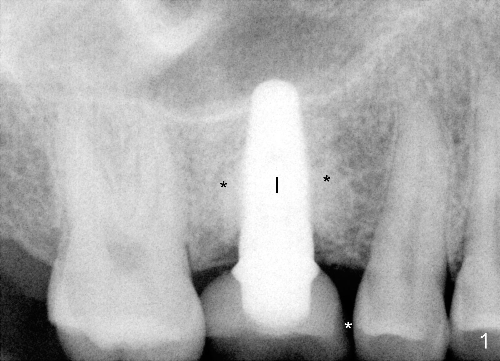
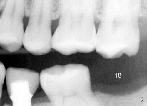
A 46-year-old man presents to clinic to fix porcelain chip over the implant crown (Fig.1 white *). The contact is loose. The patient has had history of tooth fracture, probably involving the teeth #3, 18 and 20 (Fig.2). He has habit of chewing on the right. The bone density (black *) around the implant (I) is high. All suggest bruxism.
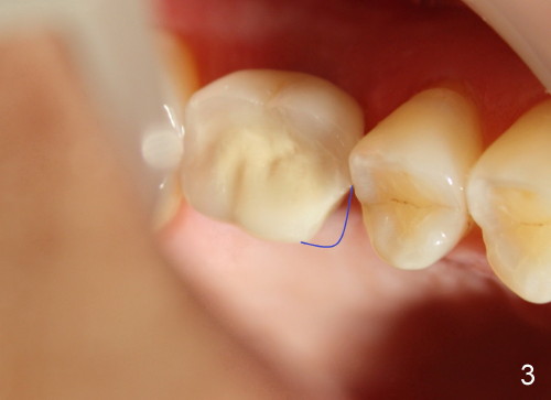
Three years after new crown fabrication, the mesiolingual cusp of the PFM fractures (Fig.3 blue outline). This time, the patient has no problem eating, since the contact is not lost. X-ray shows that the mesial portion of the porcelain is not supported by metal framework (Fig.4 ^). Our treatment plan is first to restore the missing tooth #18.
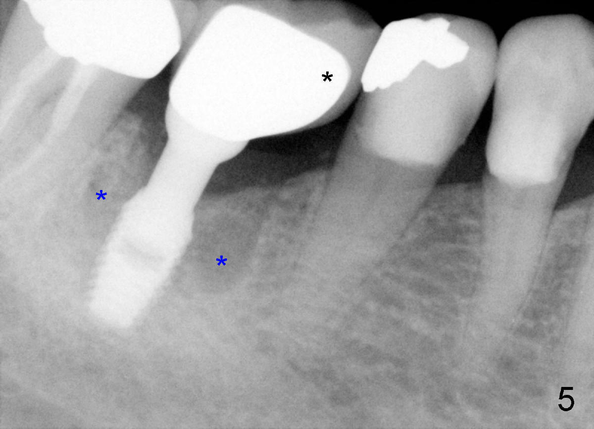
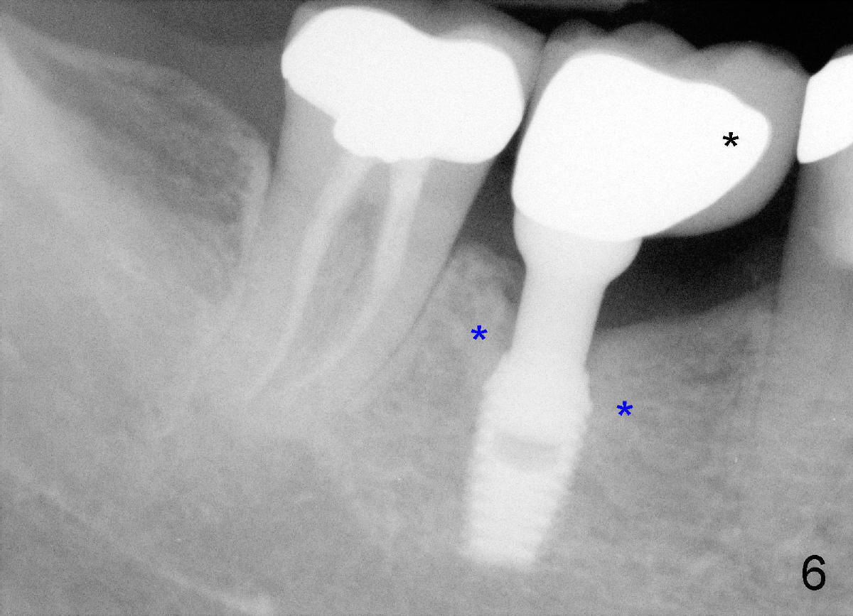
Heavy mastication tends to break not only natural dentition, but also restoration. The crown should be made of full metal, or Zirconium. Or PFM crown should be supported by larger metal framework (Fig.5 black *). This implant is placed off center (in the distal socket). Fig.5 X-ray is taken on the day of PFM cementation. Three years and a half later, there is no porcelain chip (Fig.6), while the bone density around the implant is increased (blue *), as compared to those in Fig.5. In fact the larger metal framework is designed and fabricated by a large dental lab in Florida (Smith-Sterling).
Xin Wei, DDS, PhD, MS 1st edition 09/24/2012, last revision 10/10/2012