.jpg)
|
|
|
|
|
|
|
|
|
|
||
|
|
|
|
||
|
|
|
|
|
|
Subcrestal Placement
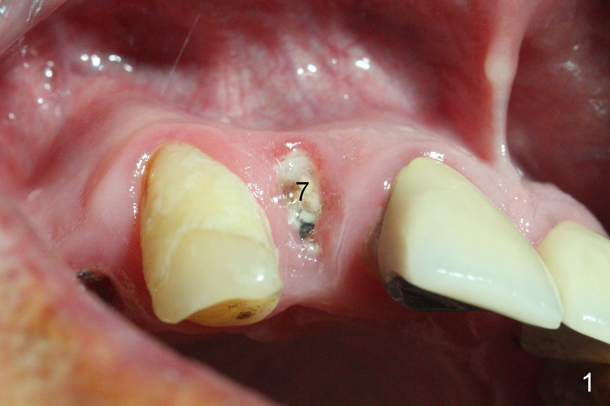
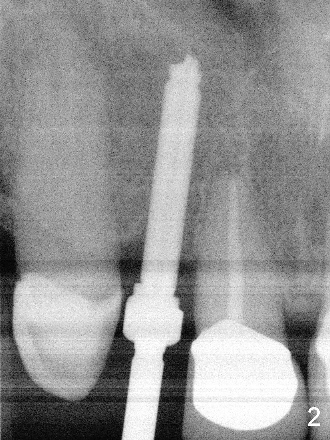
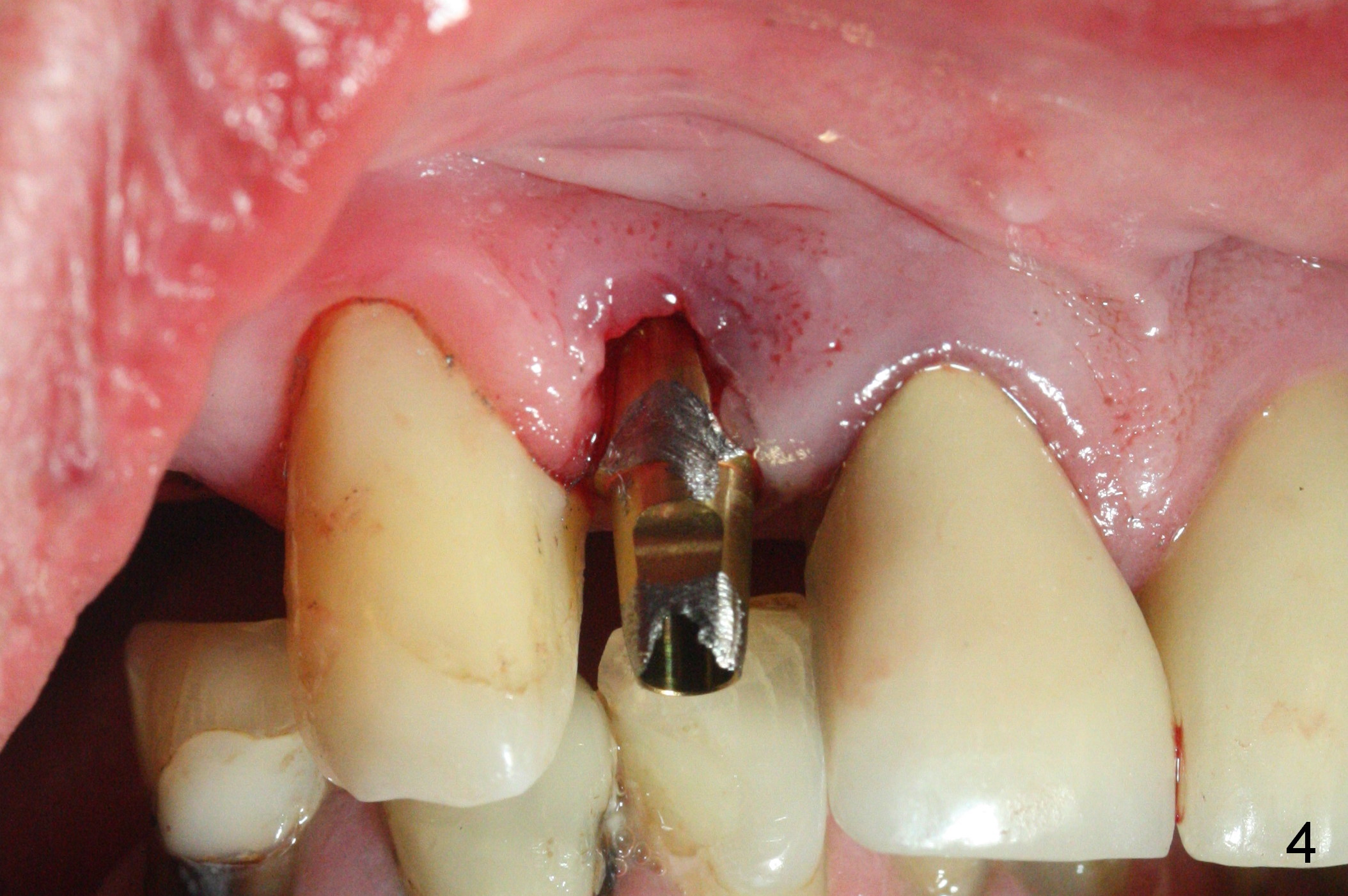
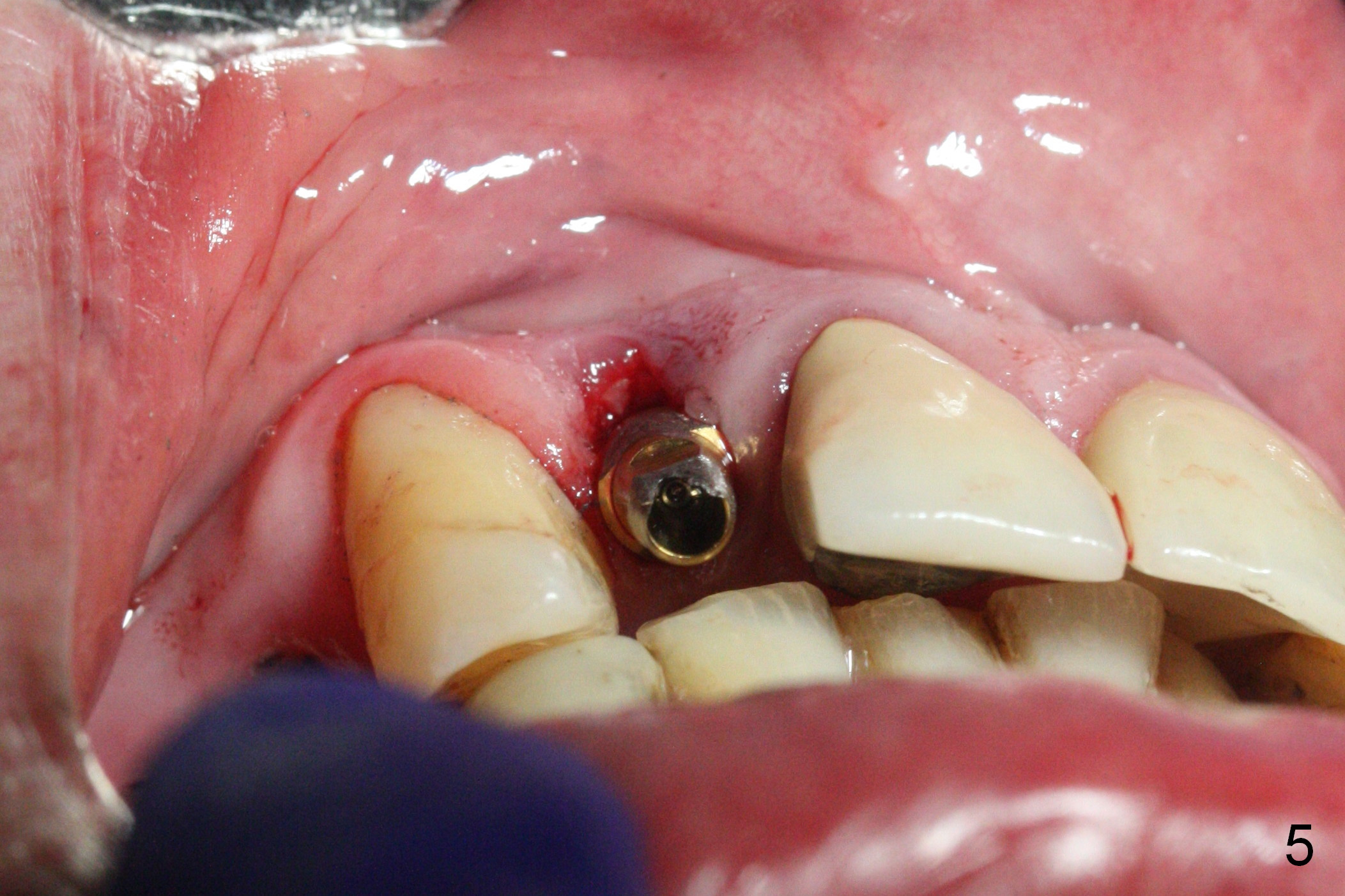
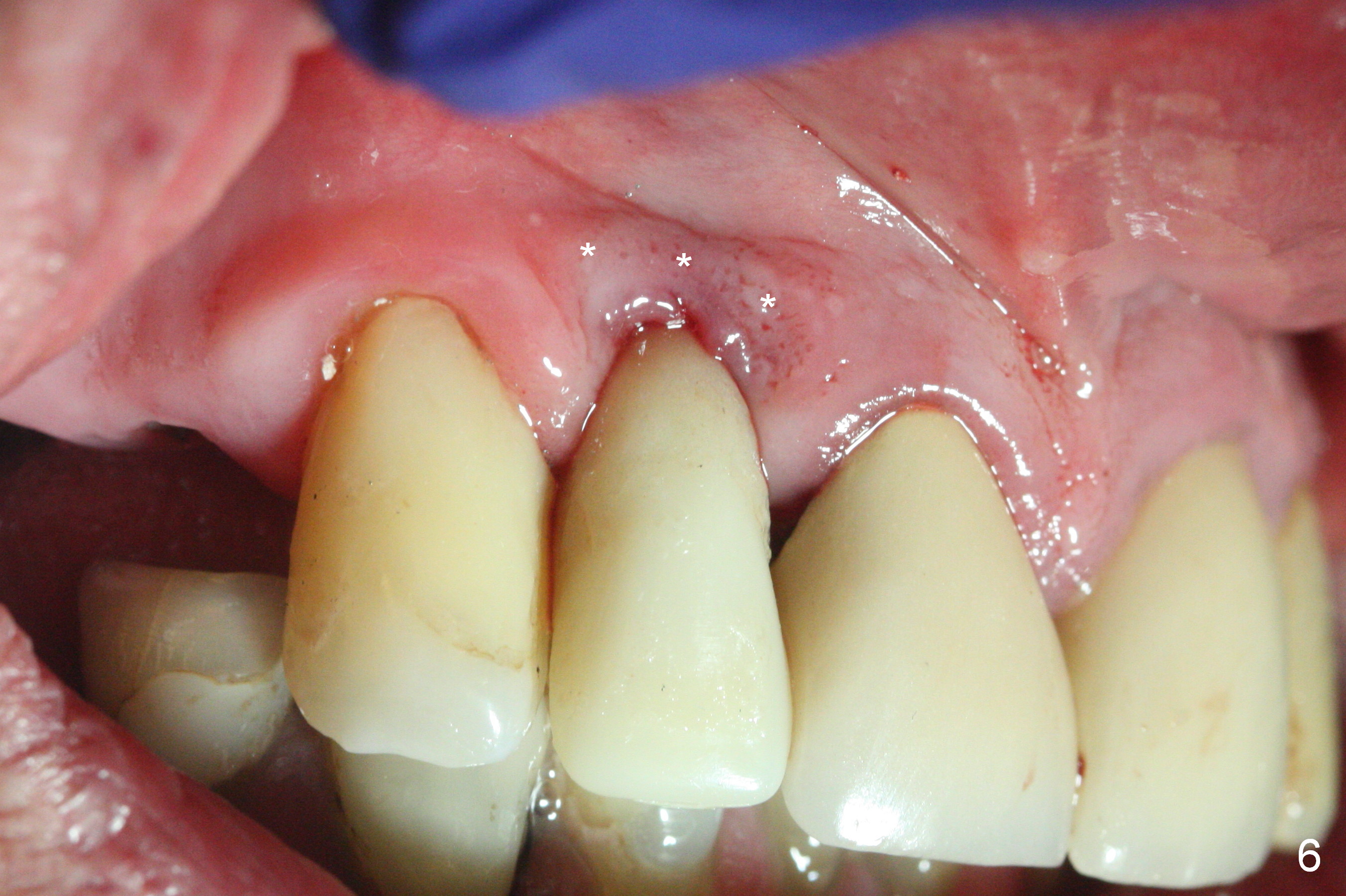
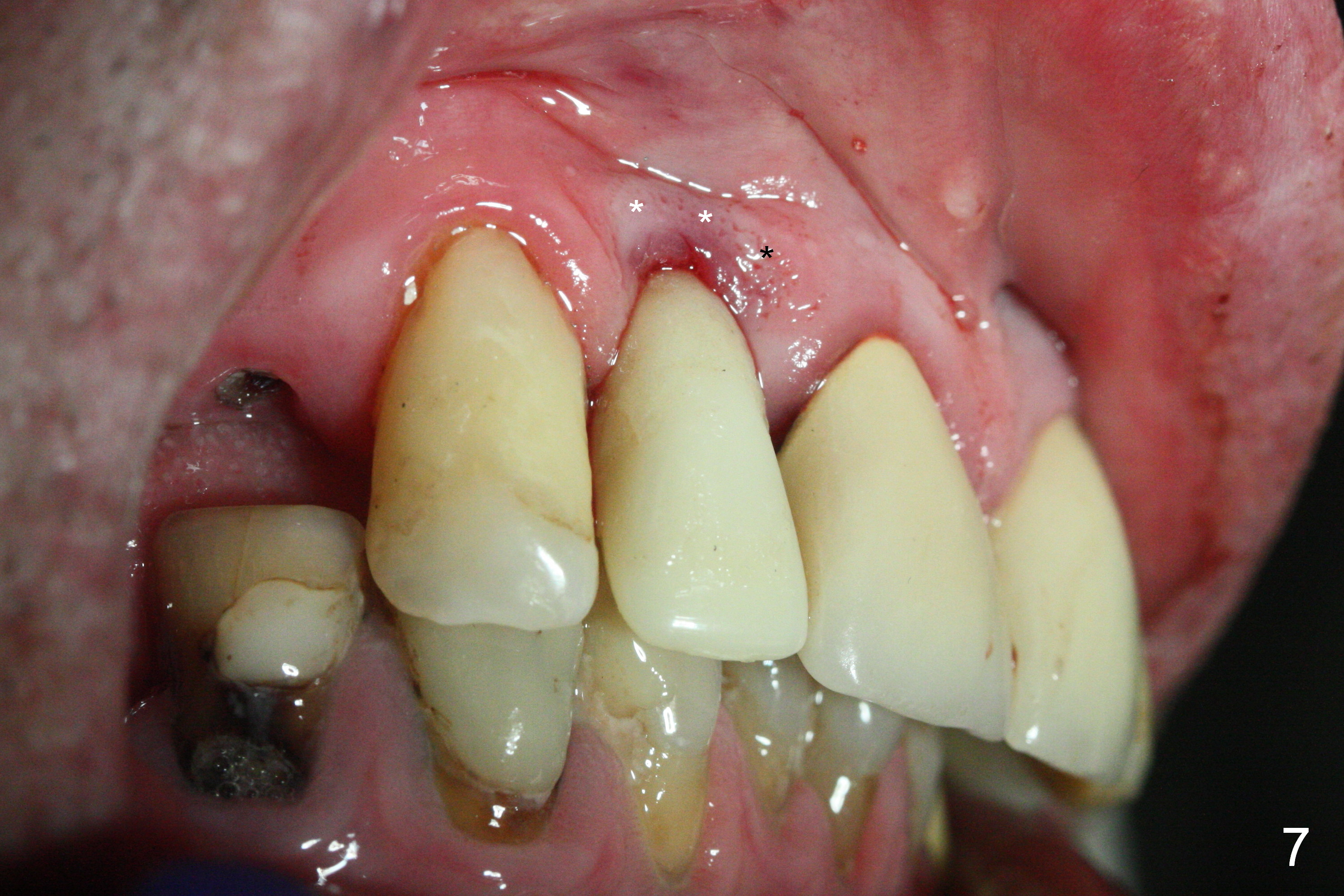
The residual root at #7 is fairly well exposed with buccal gingival recession (Fig.1). The implant placement is flapless (Fig.2-7). What is not shown is that the 3.8x13 mm implant is buccally subcrestally placed (Fig.3). After 1st round of bone graft buccal to the implant, a 4.5x5(5) mm abutment is immediately placed and prepared (Fig.4,5). An immediate provisional is placed after 2nd round of bone graft subgingivally buccally (Fig.6,7). Note the bulging gingiva (*), as compared to that in Fig.4,5. The long implant is chosen because of anterior deep bite (Fig.4) and lack of posterior support (Fig.7).
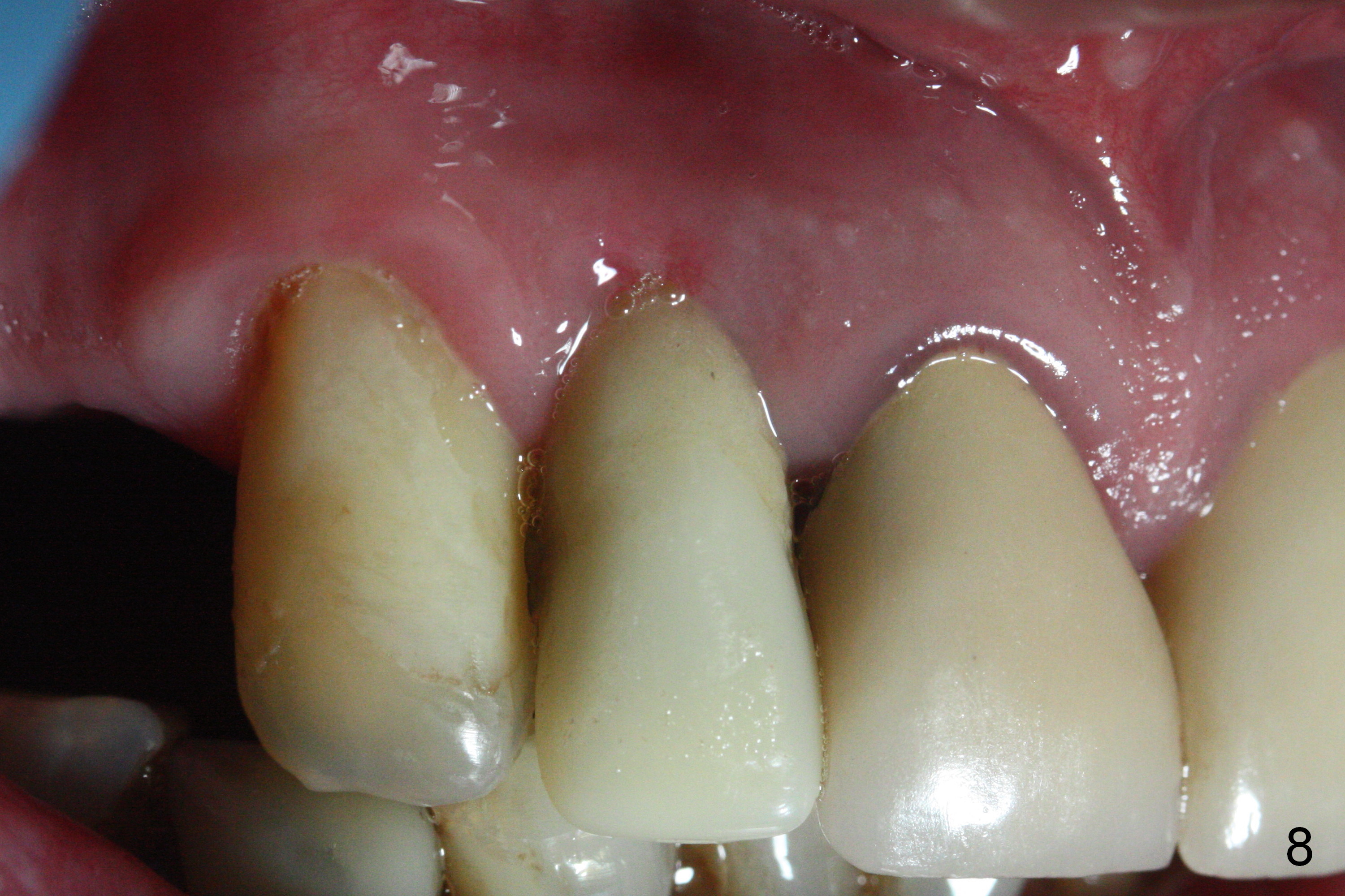
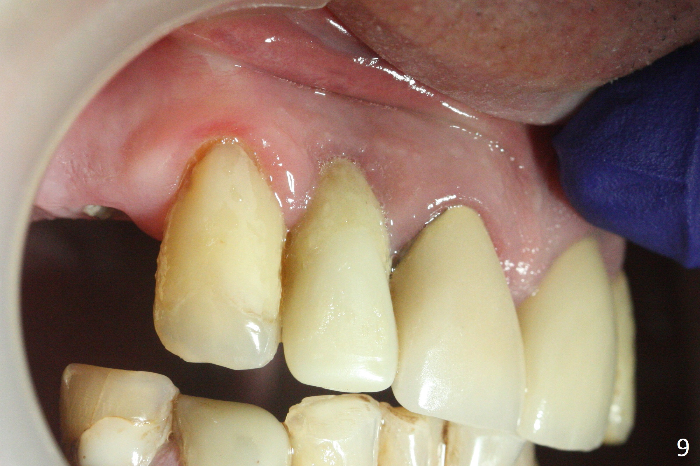
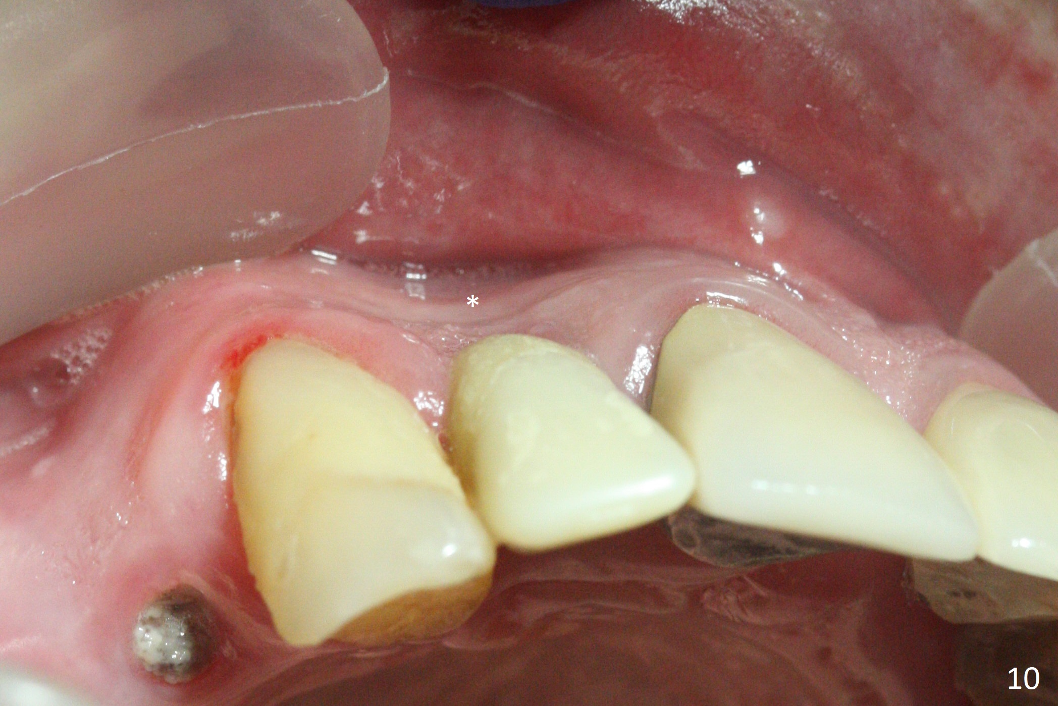
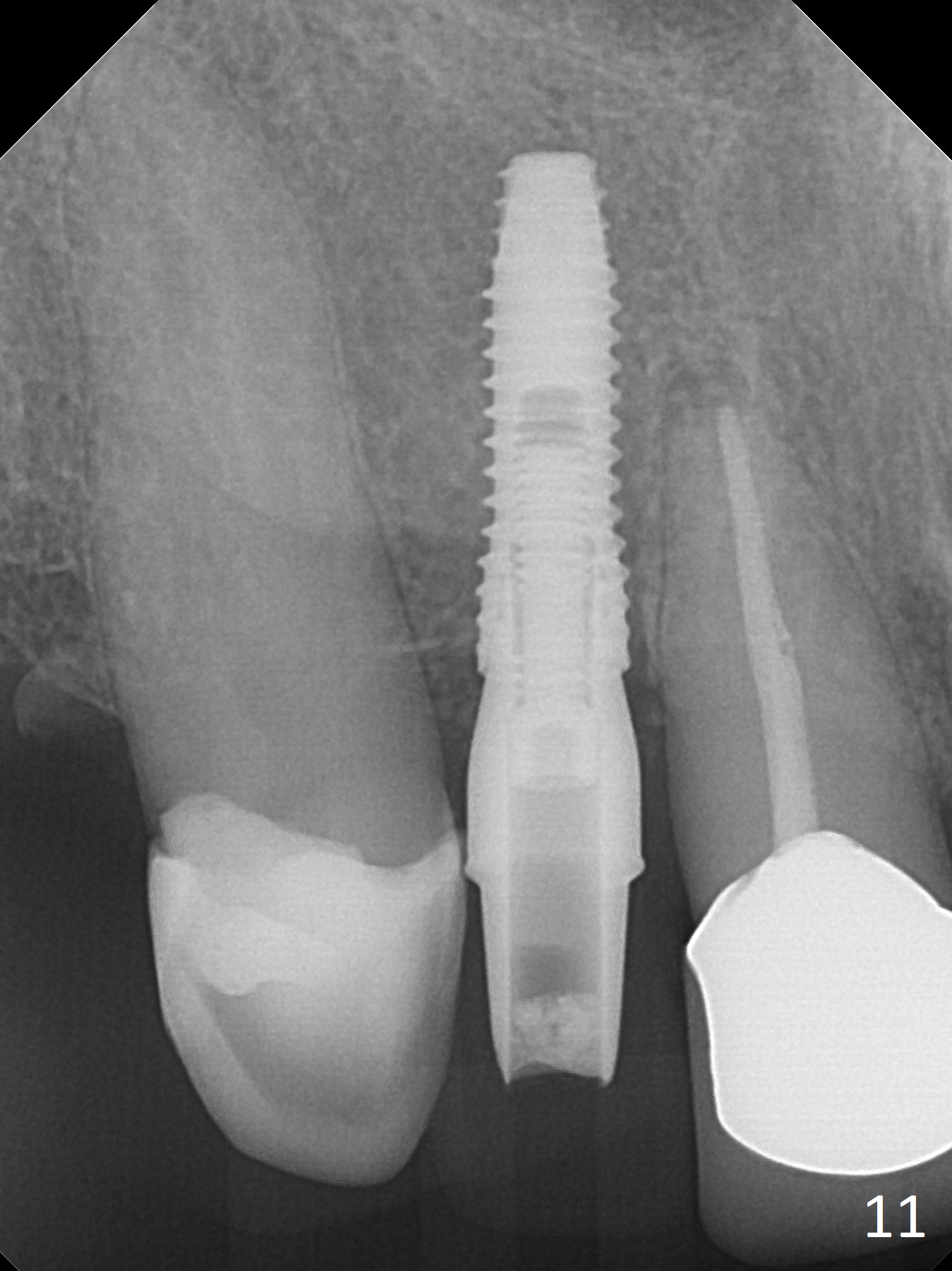
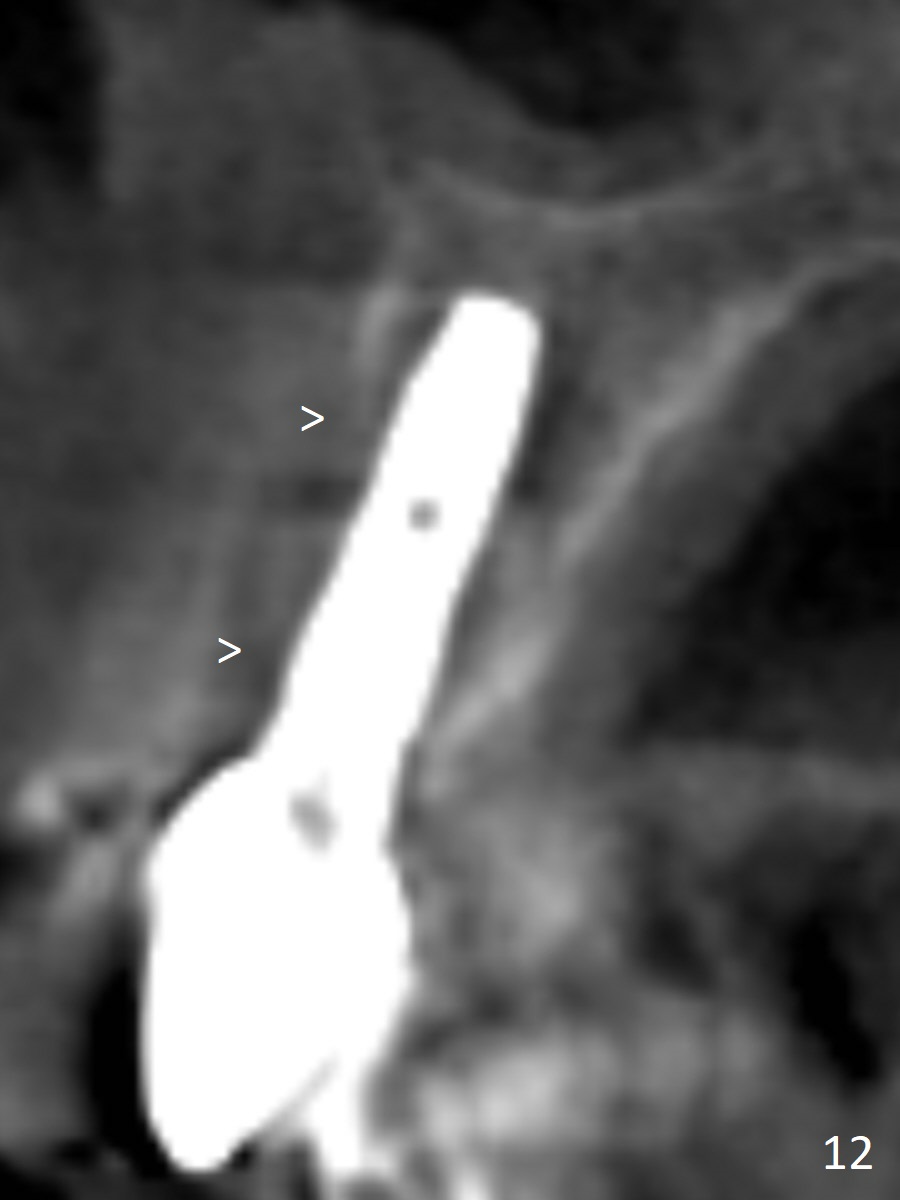
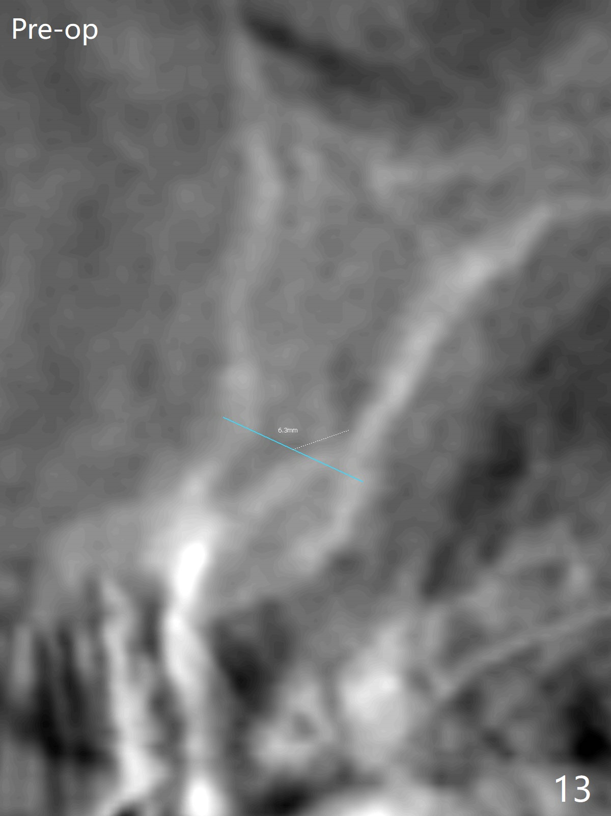
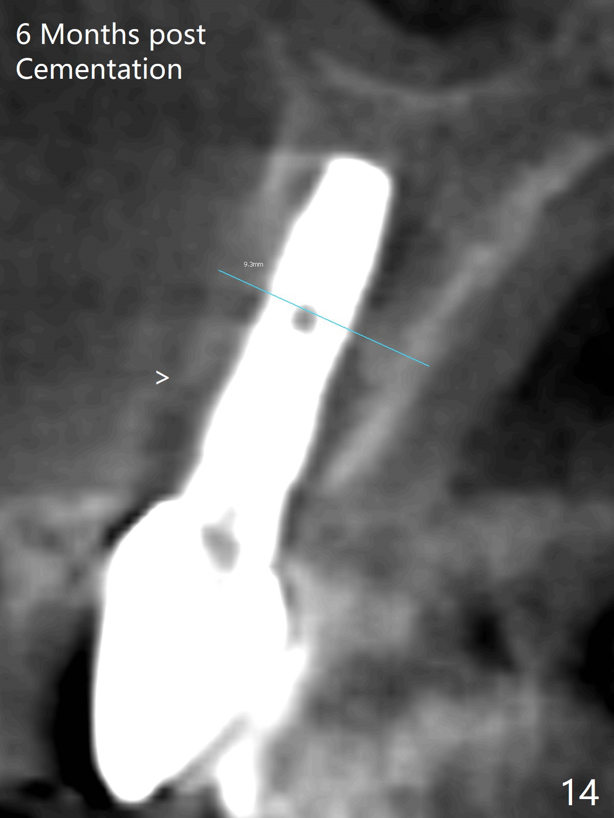
The gingiva adapts well to the provisional 1 week postop (Fig.8). The buccal gingiva remains recessive and the buccal plate is concave 5.5 months postop (Fig.9-11). It appears that the coronal portion of the buccal plate is resorbed 12 months postop (6 months post cementation, Fig.12 >). Socket shield or smaller diameter implant should have been done to avoid bone loss. Reanalysis of CBCT reveals that implantation apparently increases the bone width and that the buccal plate resorption is not so severe (Fig.13,14 (>: coronal end of the buccal plate)). Panoramic X-ray is taken 2 year 5 month post cementation.
Return to
Upper Incisor Immediate
Implant,
IBS,
Improvement, #10
22
Xin Wei, DDS, PhD, MS 1st edition 12/05/2016, last revision 11/05/2019