|
|
|
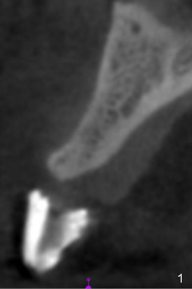
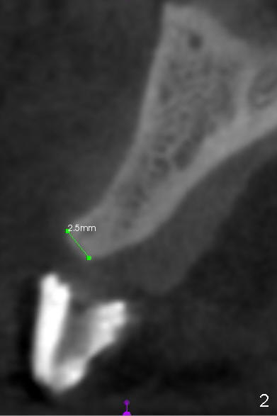
A 48-year-old lady requests fixed prosthesis at the site of #7, which is atrophic (Fig.1 CT coronal section), 2.5 mm wide (Fig.2). This CT image suggests that the bone density is moderate to high. Implant placement at the site of #14 confirms the bone density.
1 Piece or Onlay Graft? Atrophic Ridge Next Onlay
Xin Wei, DDS, PhD, MS 1st edition 11/27/2014, last revision 08/05/2018