|
|
|
|
|
|
|
|
|
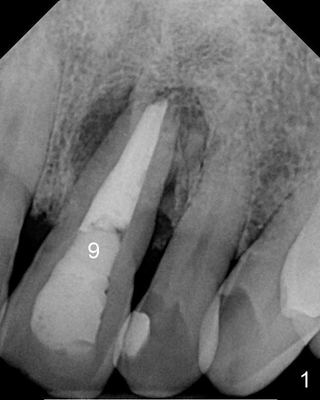
The tooth #9 has large periradicular radiolucency and sign of apicoectomy (Fig.1).
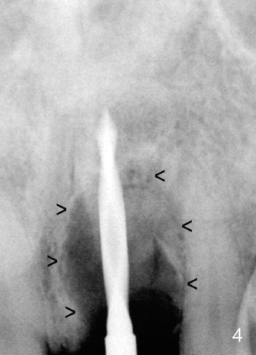
A large periradicular radiolucency (Fig.4 arrowheads) is confirmed with a 2 mm pilot drill in place (20 mm deep from the gingival margin).
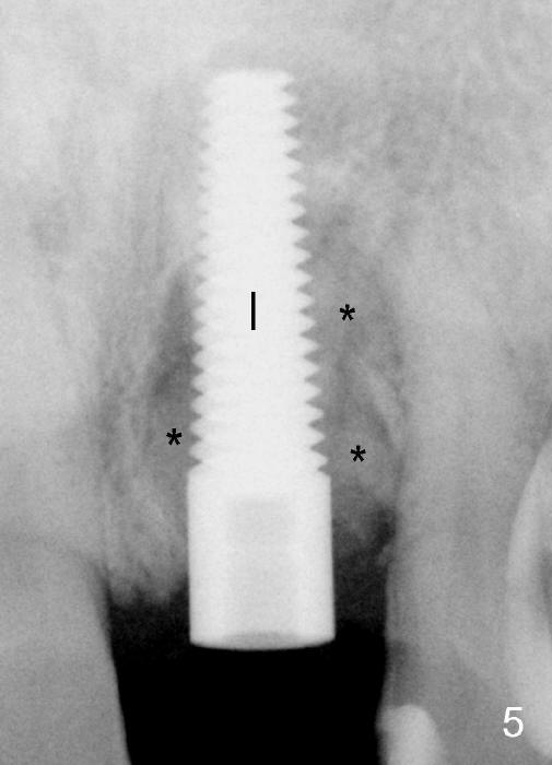
A 5x20 mm implant is placed with insertion torque >60 Ncm (Fig.5 I), with mixture of autogenous bone, allograft and synthetic bone filling the defect (*).
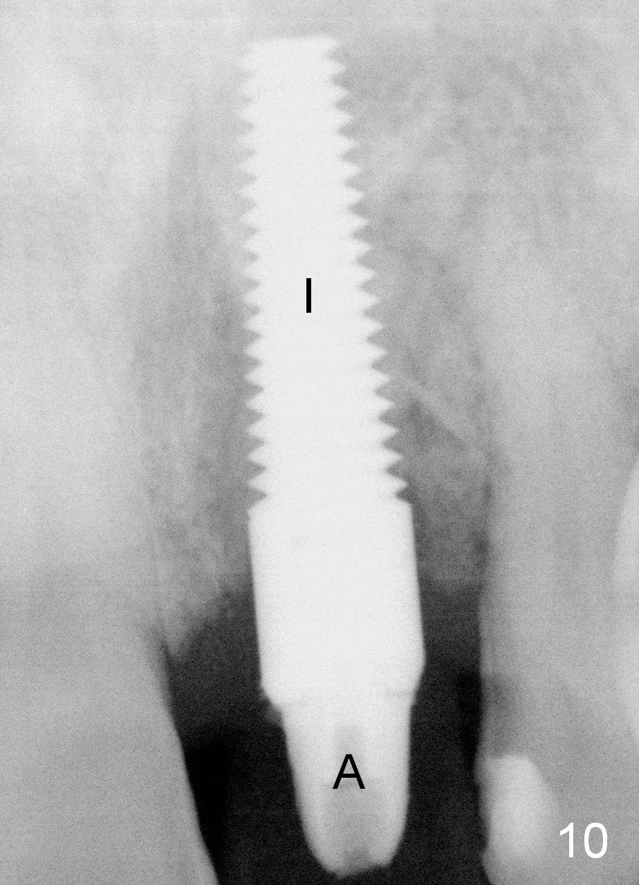
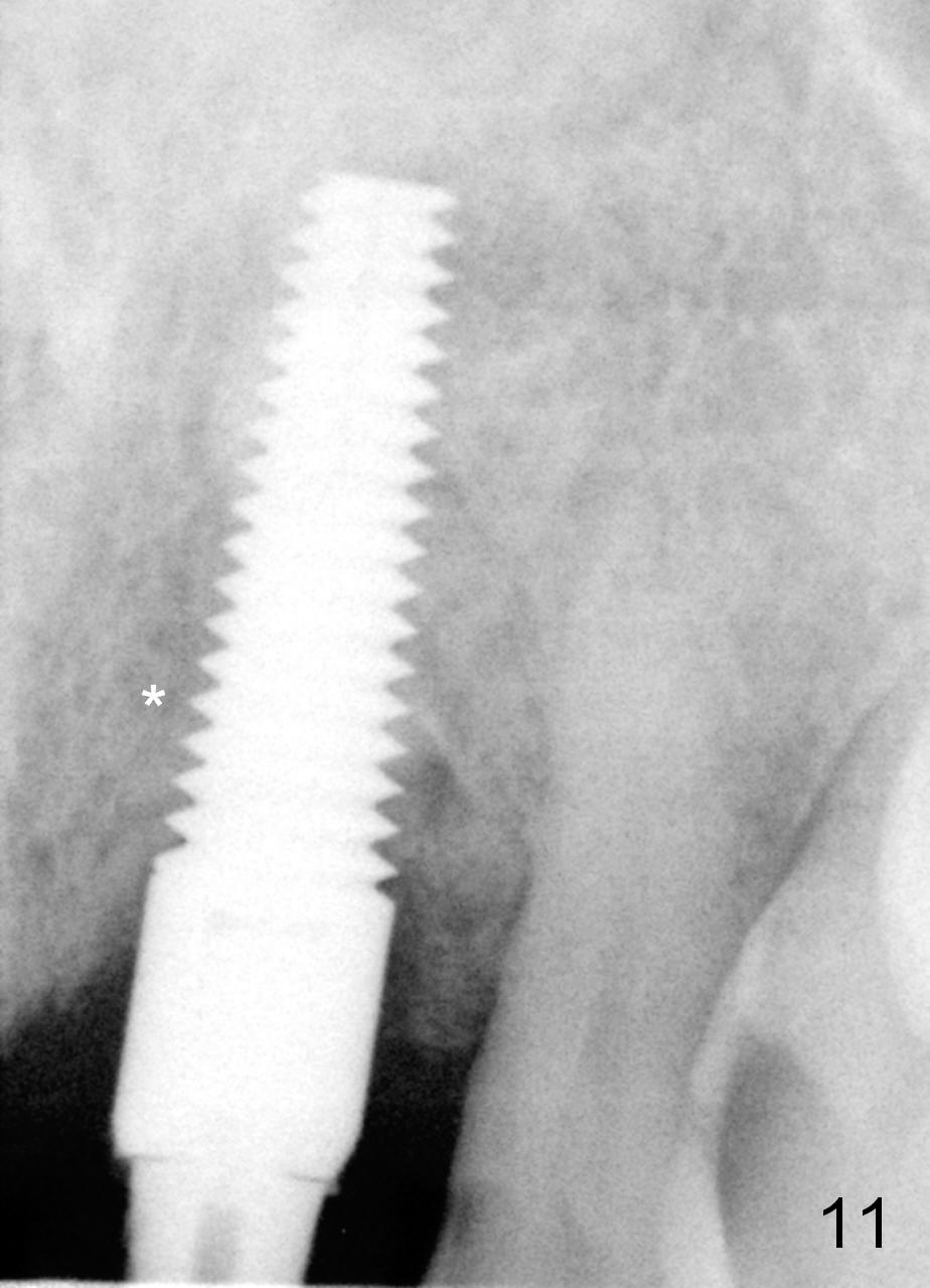
The bone density around the implant appears to increase 2 months (Fig.10 I) and 7 months (Fig.11) postop.
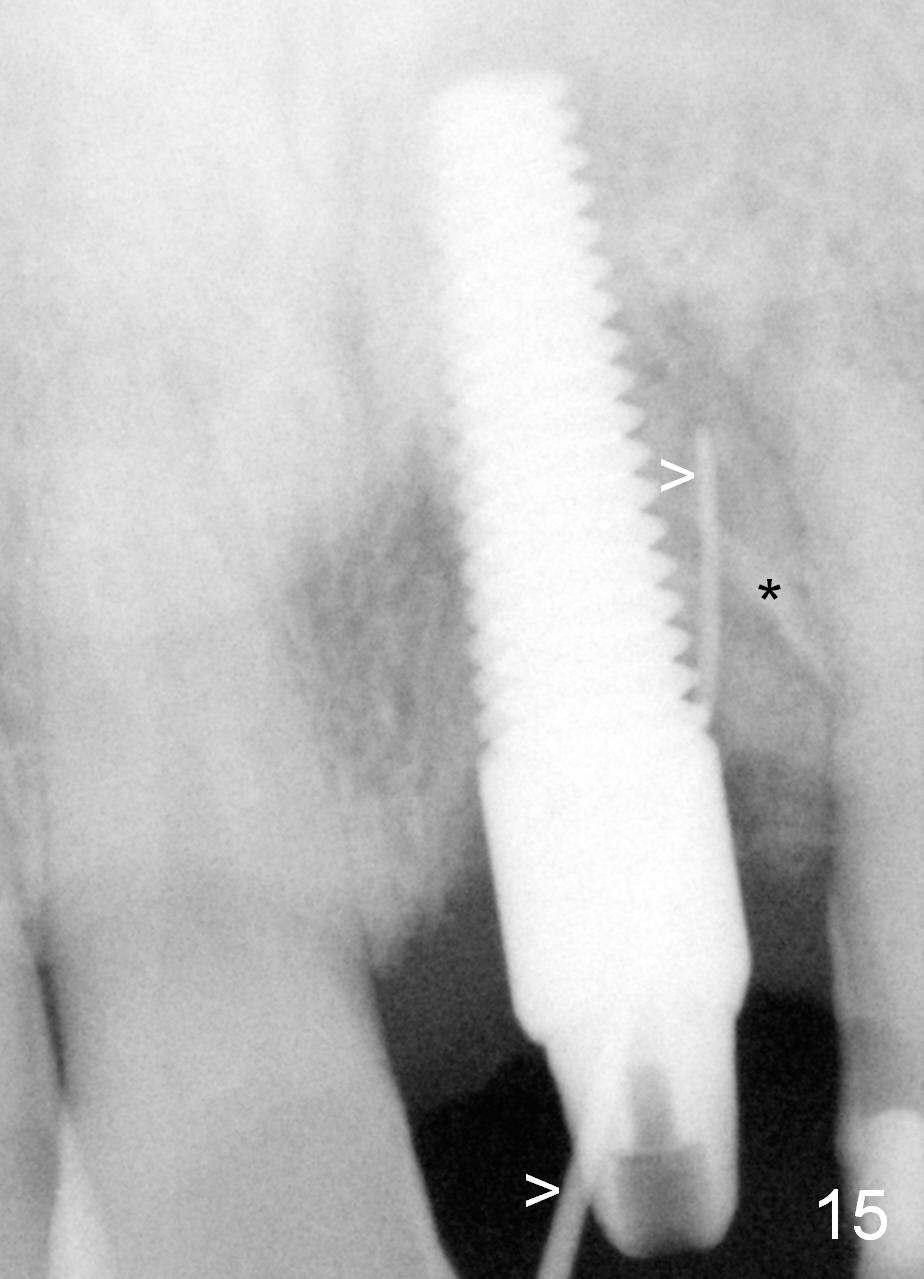
When a Gutta Percha is inserted into the fistula (Fig.15 >) before crown cementation (7.5 months postop), it points to the original mesial socket with possible foreign body (*, which may be related to apicoectomy).
How to Close Fistula Last Next
Xin Wei, DDS, PhD, MS 2/07/2014, last revision 05/27/2018