|
|
|
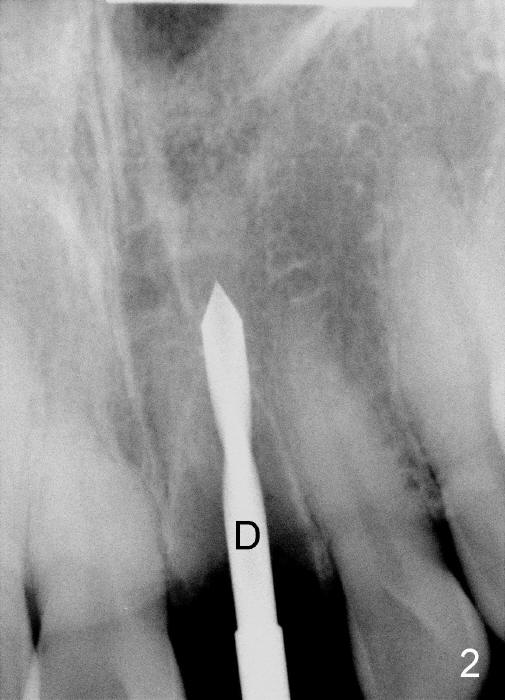
Fig.2: 2 mm pilot drill (D), close to the incisive canal.
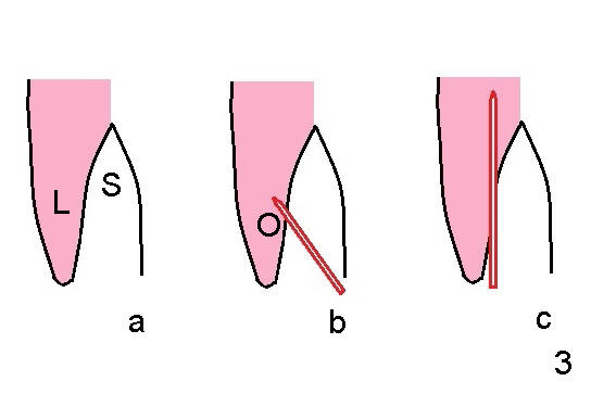
Fig.3 (illustration of sagittal section of the central incisor): Fig.3a shows the socket (S) after extraction. Fig.3b shows that initial osteotomy (O) is created by a pilot drill (red outline) in the coronal portion of the lingual plate (L) perpendicularly. Once the drill gets engaged into the lingual bone, the trajectory is changed along the long axis of the socket (Fig.3c).
Return to Osteotomes Last Next
Xin Wei, DDS, PhD, MS 1st edition 06/19/2013, last revision 06/23/2014