|
|
|
|
|
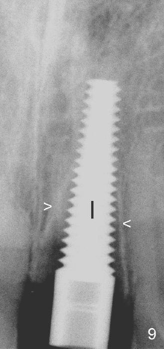
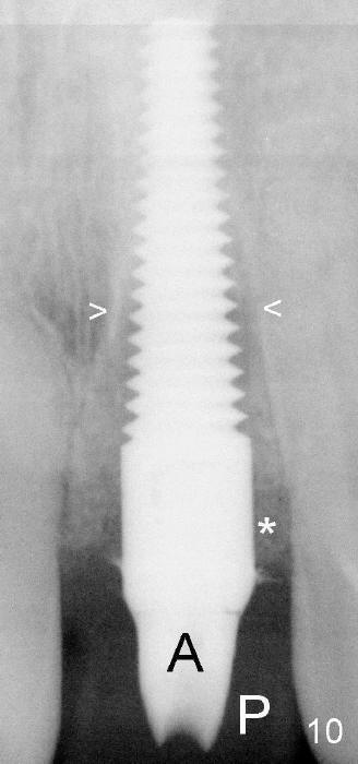
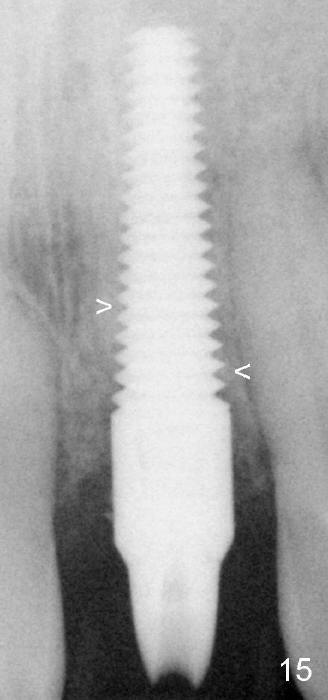
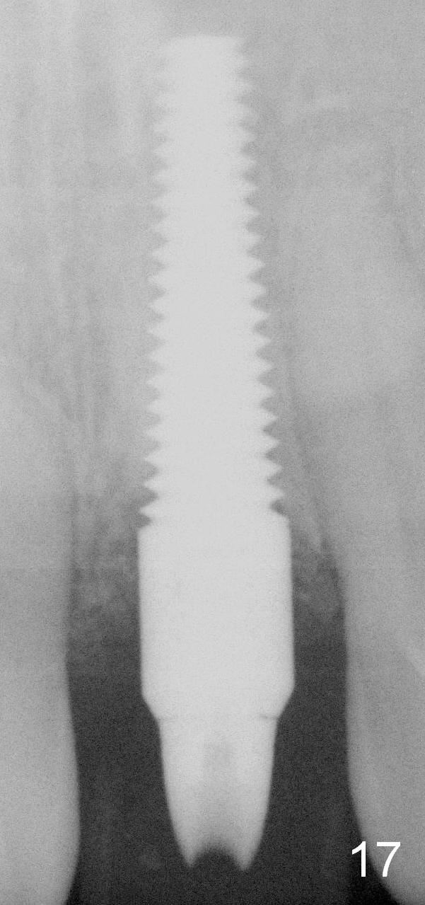
When a 4.5x20 mm implant (Fig.9 I) is placed and allograft fills the coronal peri-implant space (Fig.10 *), the lamina dura is distinct (arrowheads). Three months postop, the lamina dura is blurred, while new bone appears to have grown into the coronal threads of the implant (arrowheads). (A: abutment; P: provisional). Osteointegration is more obvious 9 months postop (Fig.17).
Return to Osteotomes Last Next Anatomy
Xin Wei, DDS, PhD, MS 1st edition 06/19/2013, last revision 04/02/2015