|
|
|
|
|
X-ray for Upper Canine
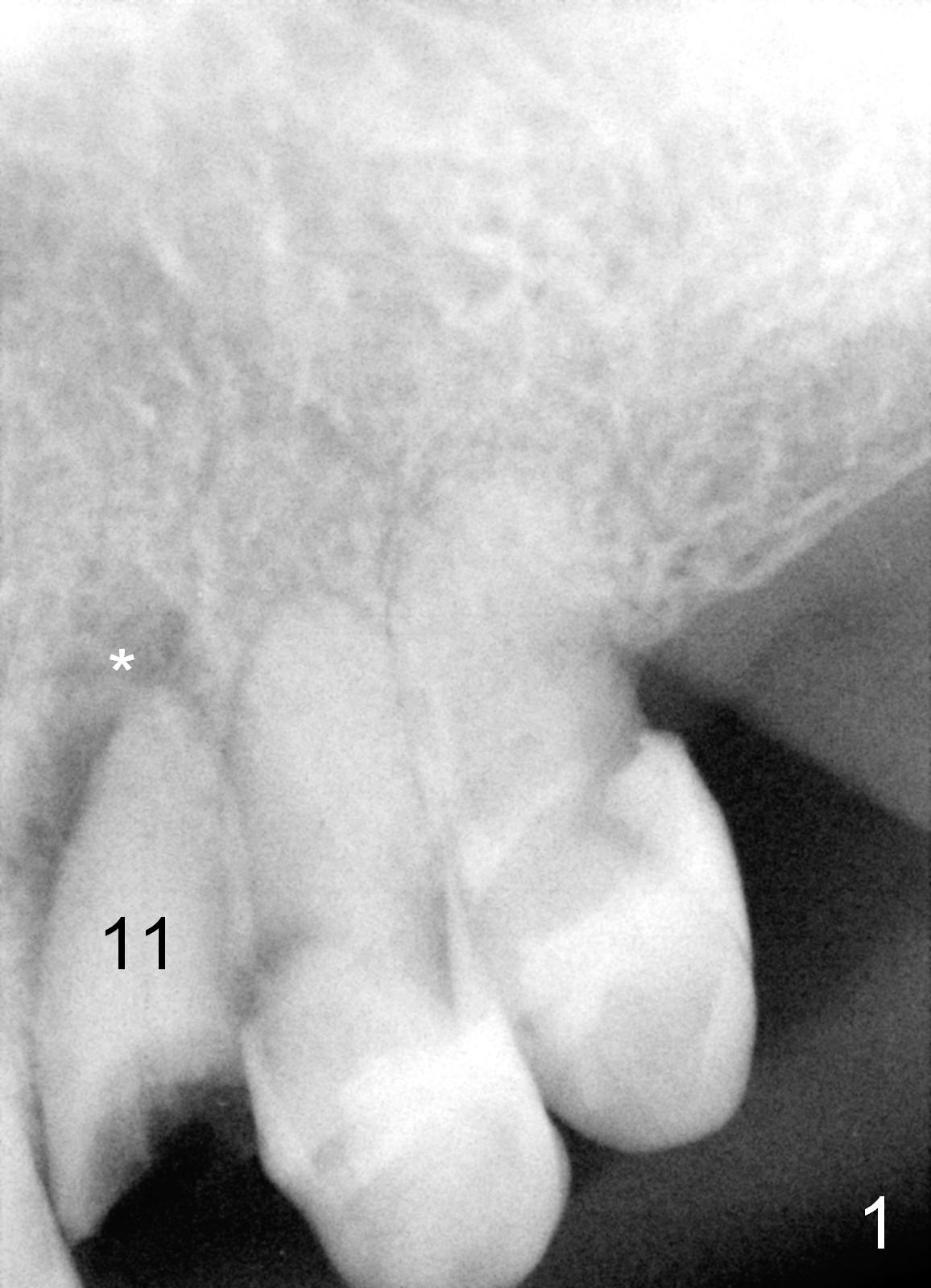
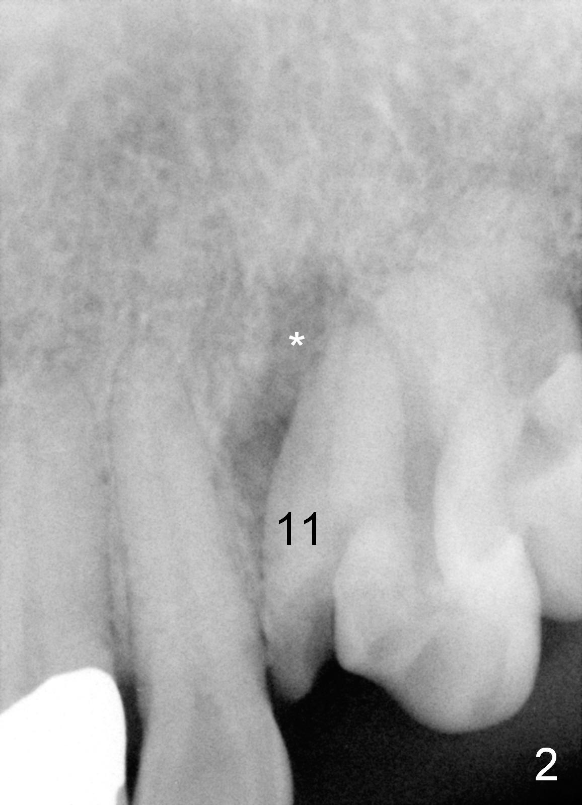
The upper canine is at the corner of the arch. Taking X-ray for the canine is tricky. The senor should be placed at the right position and X-ray cone should be placed at the right angle. For example, PA sensor in Fig.1 is placed too distal, while that in Fig.2 is placed at the right position, but the X-ray tubing is not placed at the right (correct) angle (not perpendicular to the sensor).
A 74-year-old man fractures his upper left canine (Fig.1,2: #11). The tooth has large periapical radiolucency (*).
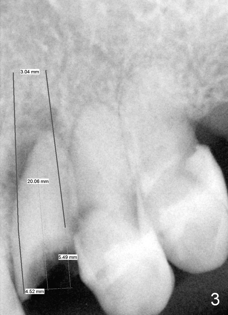
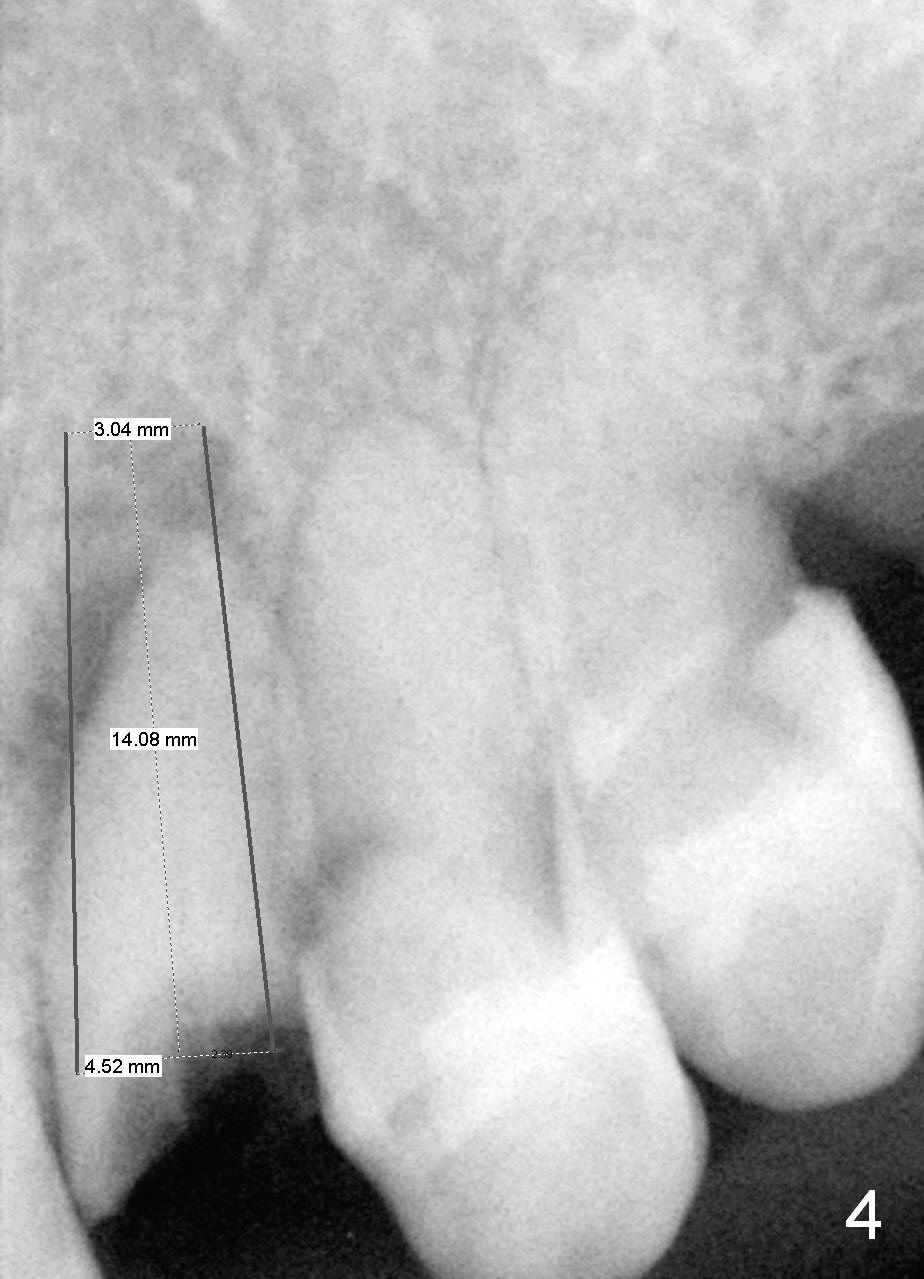
To achieve primary stability, it is more likely to place a longer tissue-level implant (Fig.3) than bone-level one (Fig.4).
Prepare a surgical handpiece in case of sectioning.
Take photos to show the root stump, caries, and buccal plate thickness. Also prepare Boley gauge to measure root diameter in reference to implant diameter.
Return to Upper
Canine Immediate Implant
Xin Wei, DDS, PhD, MS 1st edition 07/01/2015, last revision 02/03/2019