|
|
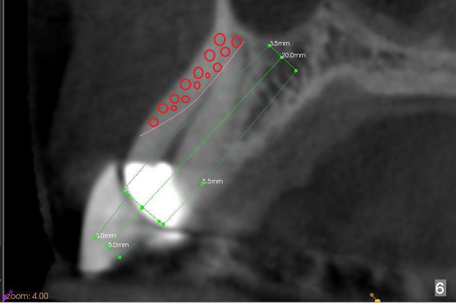
Since the canine socket is large, particulate bone graft is placed in the buccoapical area of the socket (Fig.6 red circles) prior to implant placement. A curette is used to pack the graft against the labial wall and there is no blockage to the opening of the osteotomy. More bone graft is placed buccal and coronal to the implant once the latter is placed.
CT for Planning Last Next
Xin Wei, DDS, PhD, MS 1st edition 11/28/2014, last revision 02/03/2019