|
|
|
|
|
|
|
|
Repair of Buccal Plate Associated with Implant Failure
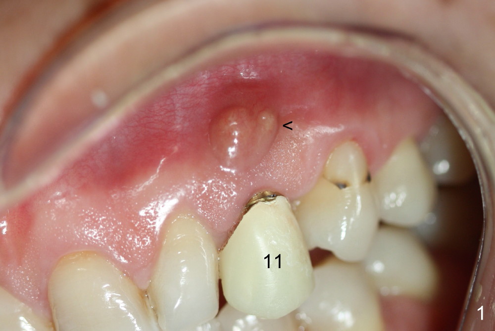
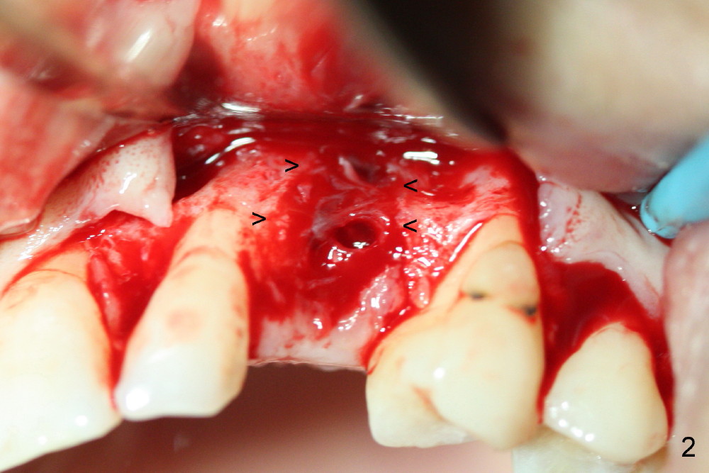
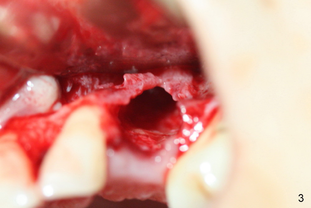
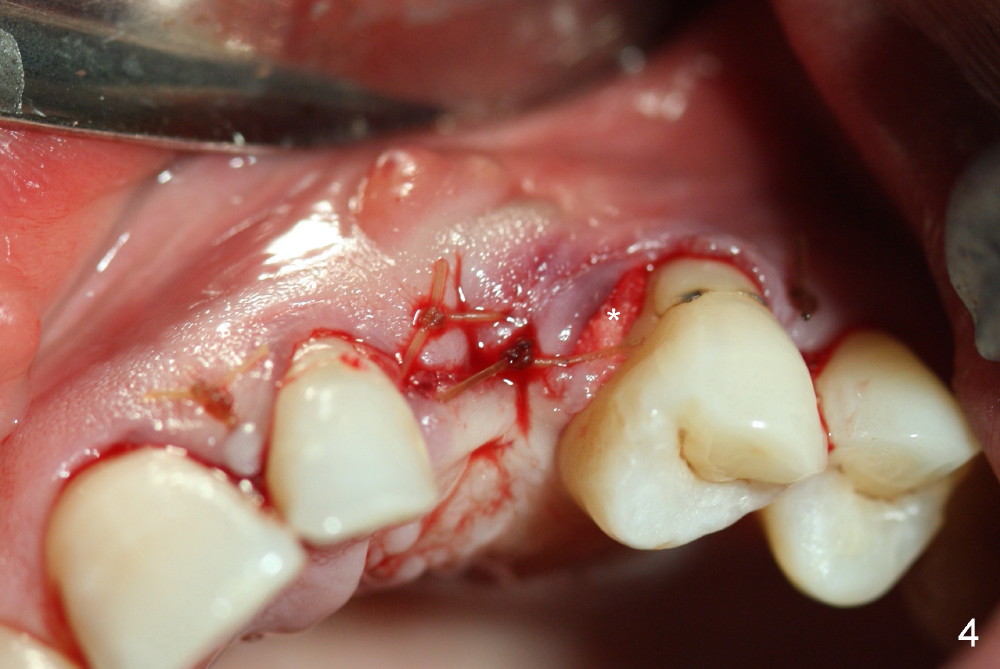
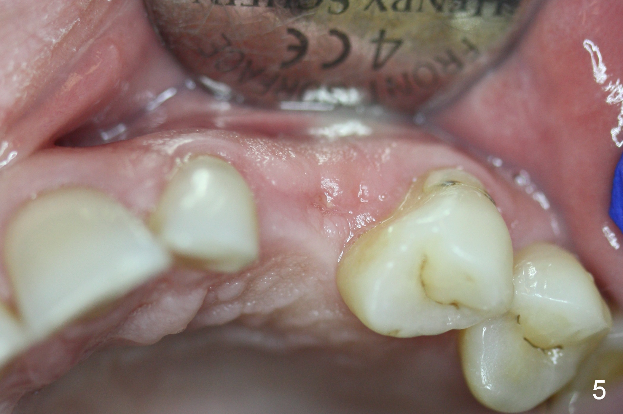
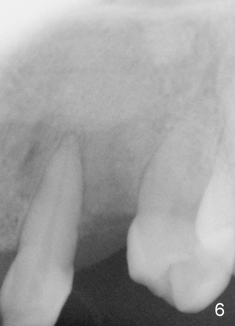
One month post-implantation at the site of the upper left deciduous canine, the patient returns for follow-up with chief complaint "The crown is a little loose. A bubble is becoming smaller". Exam shows an abscess (Fig.1 <) above the provisional (#11). The provisional is loose with mild tenderness. A pull on the provisional leads to dislodgement of the abutment and implant. Raising the buccal flap reveals perforation of the buccal plate with granulation tissue (Fig.2). After debridement (Fig.3), allograft and Osteogen is placed in the defect, followed by Osteotape (Fig.4 *) and suture. This complication can be prevented by CT information. A deciduous tooth socket is small. The immediate implant should be not too large, leaving 2 mm buccal gap. Three months 20 days post graft, the ridge looks not so atrophic (Fig.5). There is no sign of bone resorption (Fig.6). The patient will return for implant placement for the second time soon. Measure the bone width with bone caliper after local anesthesia. Fully expose the alveolus. Start osteotomy as palatal as possible. Repeatedly check osteotomy position and trajectory. Use surgical stent. The implant will not be too long or too wide. Is the ridge as wide as clinically shown?
Return to Upper Canine Immediate Implant Xin Wei, DDS, PhD, MS 1st edition 11/24/2014, last revision 02/03/2019