|
|
|
|
||
|
|
|
|
|
|
|
|
|
|||
Off Axis Implant
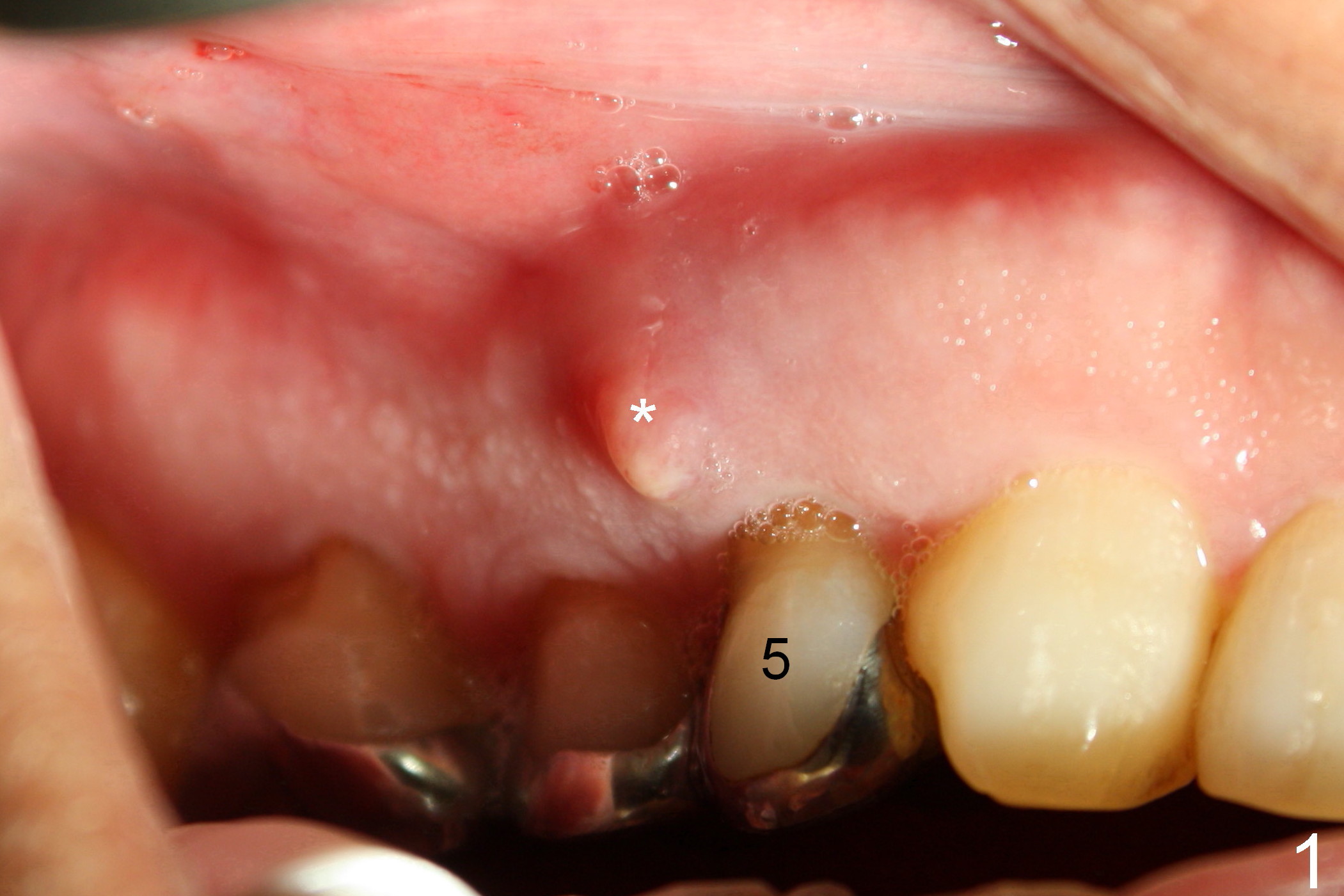
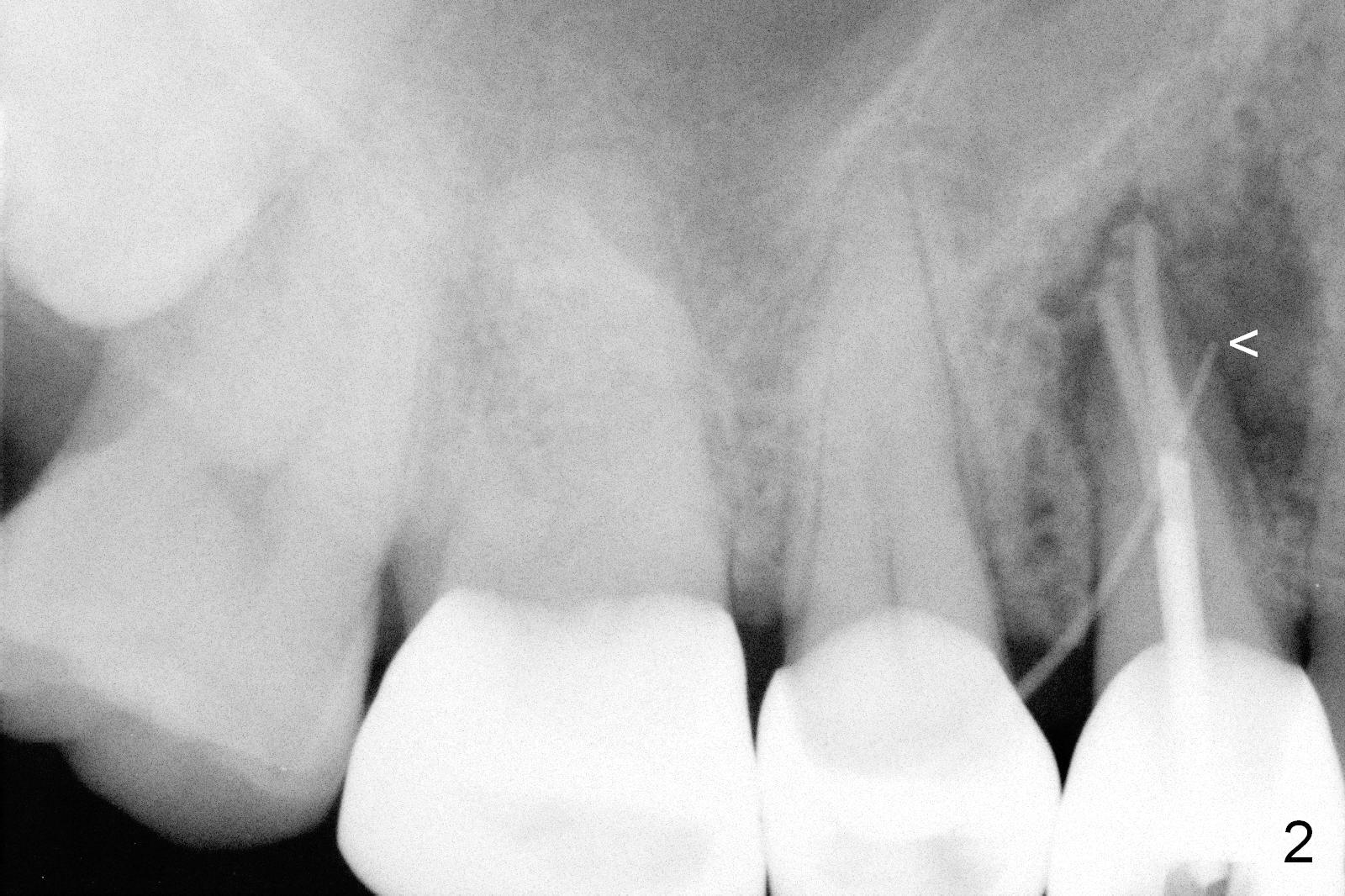
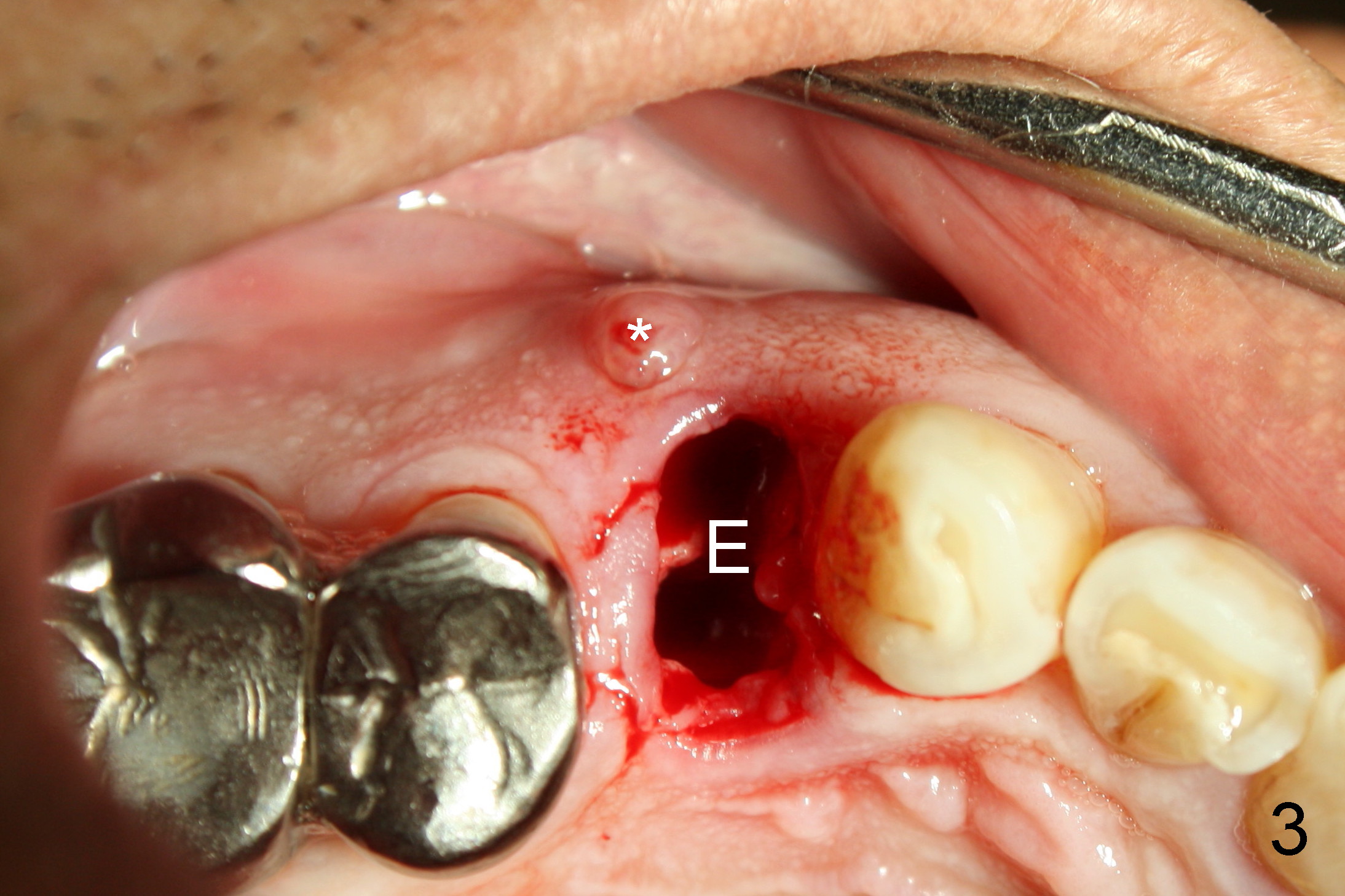
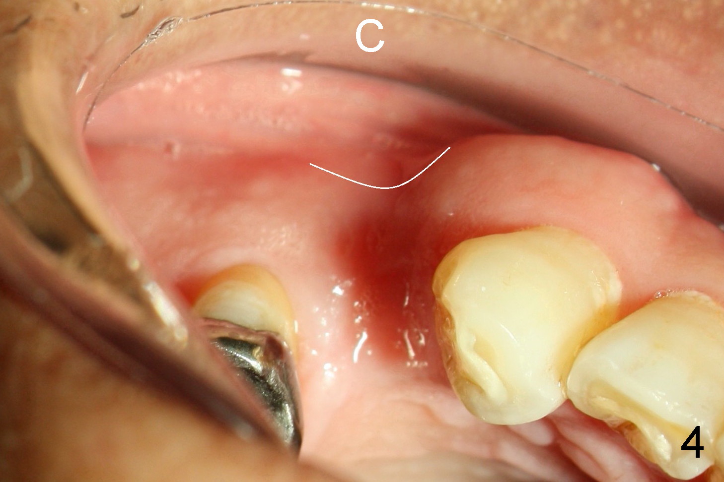
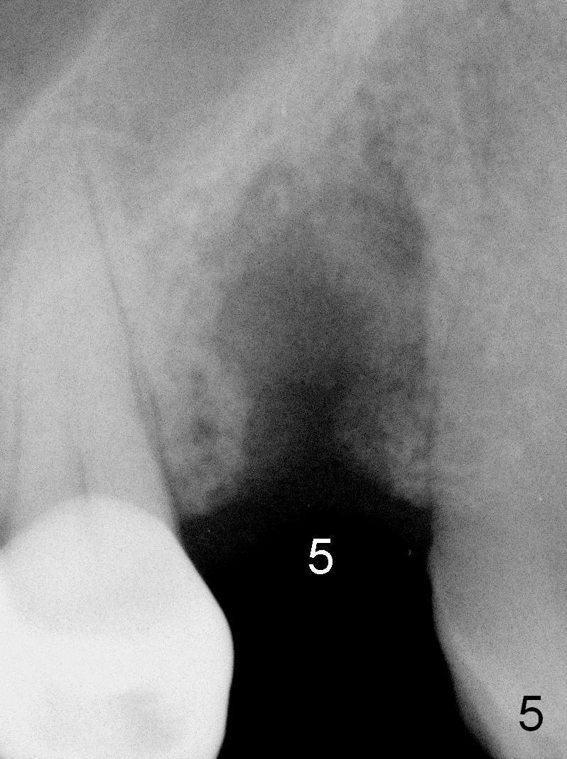
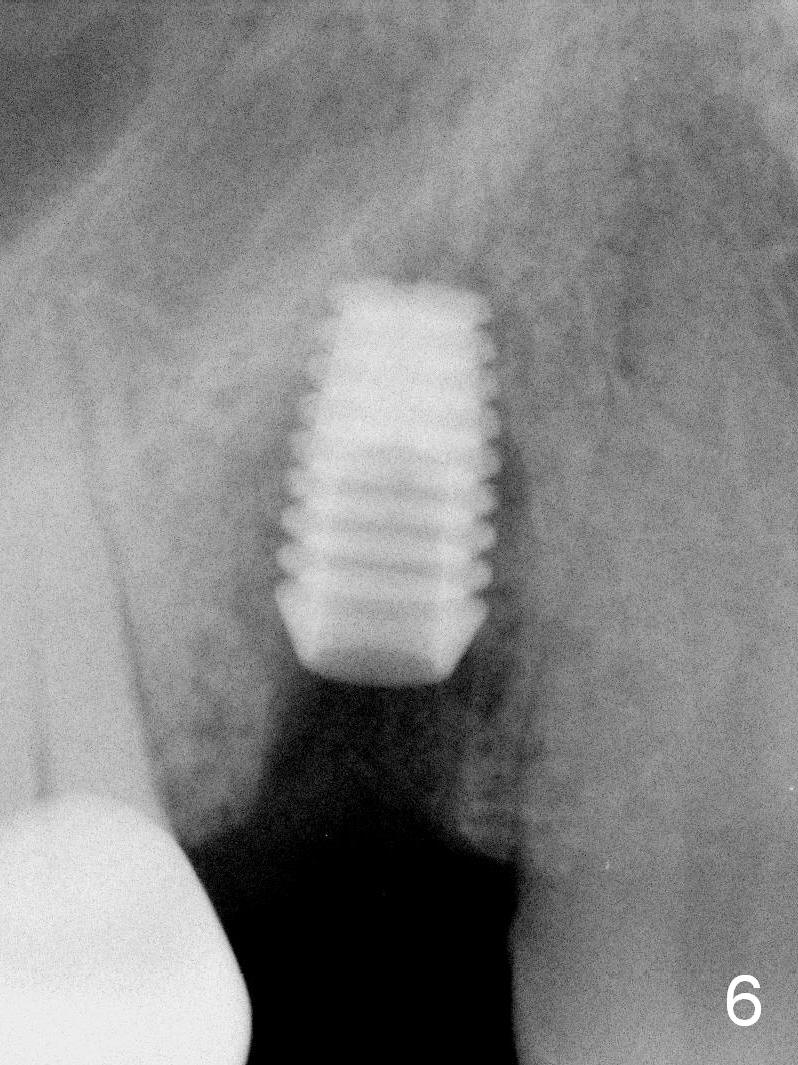
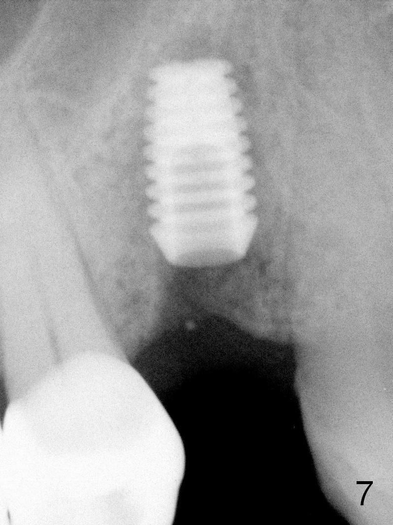
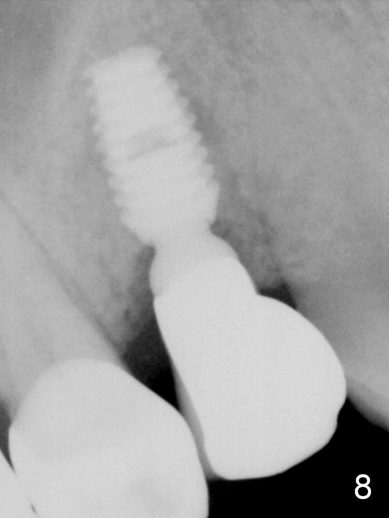
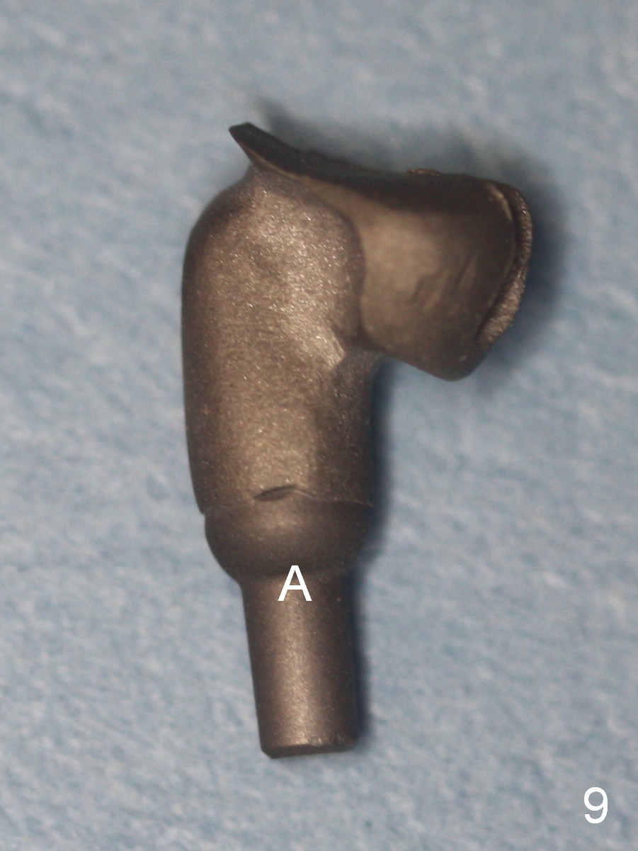
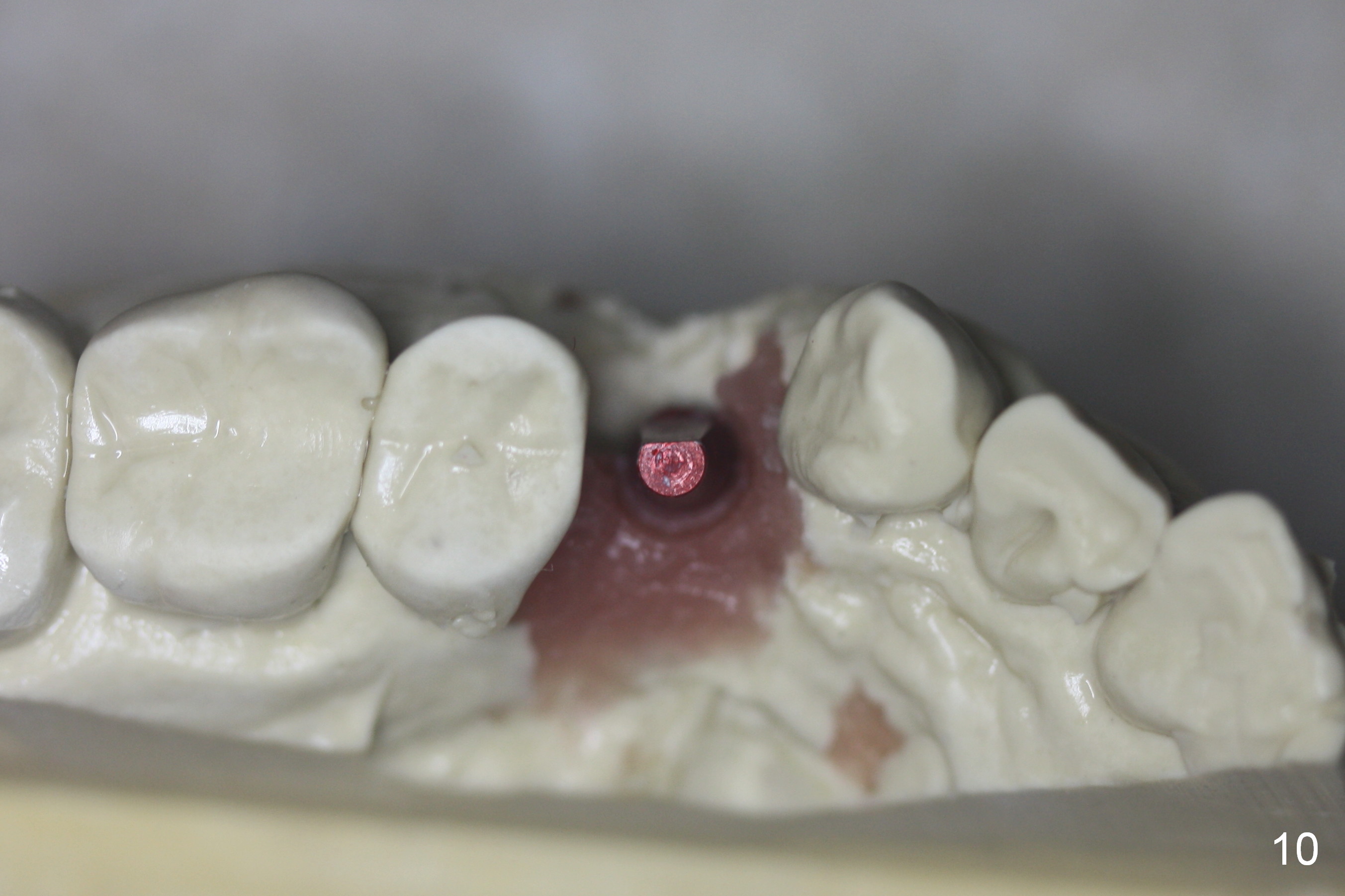
A 68-year-old man has pain and swelling associated with the tooth #5 (Fig.1). The fistula (*) is connected to the periapical radiolucency using a gutta percha (Fig.2 <). There are deep pockets distobuccal and lingual. The lingual root is found to have oblique fracture upon extraction. The distobuccal plate perforates (Fig.3). Collagen plug is placed. The buccal plate is concave (Fig.4) and socket density is low 2.5 months post extraction. Three months post extraction a 4.5x8 mm Bicon implant is placed after reamer and osteotome osteotomy (Fig.6). Bone density around the implant appears to increase 5 months post placement (Fig.7). Porcelain-fused-to-metal crown is cemented 2 weeks later. Bucco-occlusal porcelain chips 2 months post cementation. The patient decides to redo the crown. PA is taken before crown removal (Fig.8: 3 year 10 months post cementation). When a straight abutment is removed (Fig.9 A), a 15° angled abutment has to be used (Fig.10 red) for restoration.
Return to Upper Premolar Immediate Implant,
Long-term Follow Up
Xin Wei, DDS, PhD, MS 1st edition 04/17/2016, last revision 05/19/2018