|
|
|
|
|
|
|
|
Poor Man CT for Upper Molar Immediate Implant
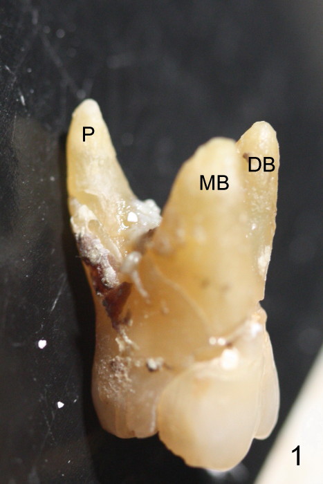
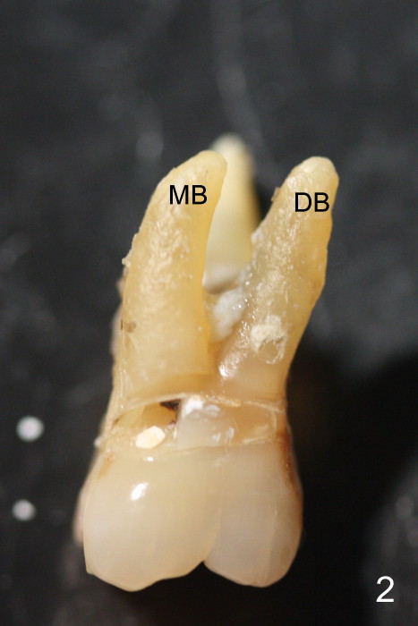
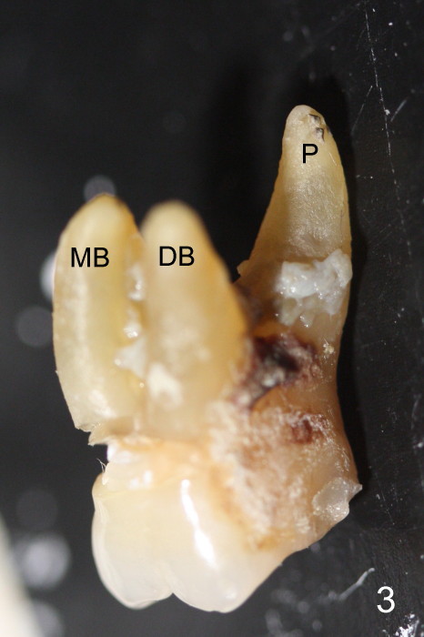
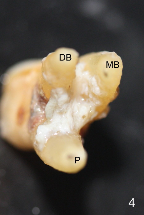
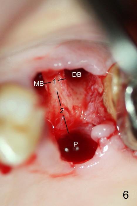
The upper 1st molar has 3 furcated roots (Fig.1-4, extracted tooth), between which is the septum (Fig.6). The septum is one of potential immediate implant placement sites, probably the best one. It would be nice to get preop 3-D septal morphology handily.
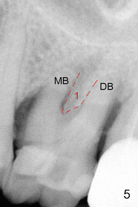
Routine straight PA shows the portion of the septum between the buccal roots (Fig.5: #1, corresponding to #1 in Fig.6). This portion of the septum is usually not the ideal implant site.
In contrast, the length of #2 (buccopalatally) in Fig.6 is critical to implant osteotomy. This piece of information is easily obtained when X-ray is taken obliquely (sideway, e.g., Fig.1,3). As well known, the septum looks like an inverted pyramid (Fig.4).
Taking 3 PAs in different angulations (Fig.1-3) can produce 3-D information in a much lower radiation dose, as compared to Cone beam CT. This is more important when a 2nd upper molar (with less root furcaton) needs implant placement.
Return to Upper Molar Immediate Implant
Xin Wei, DDS, PhD, MS 1st edition 12/26/2014, last revision 12/26/2014