|
|
|
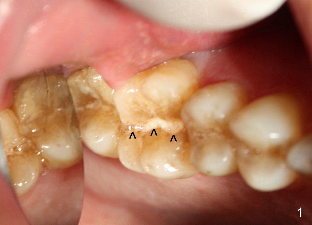
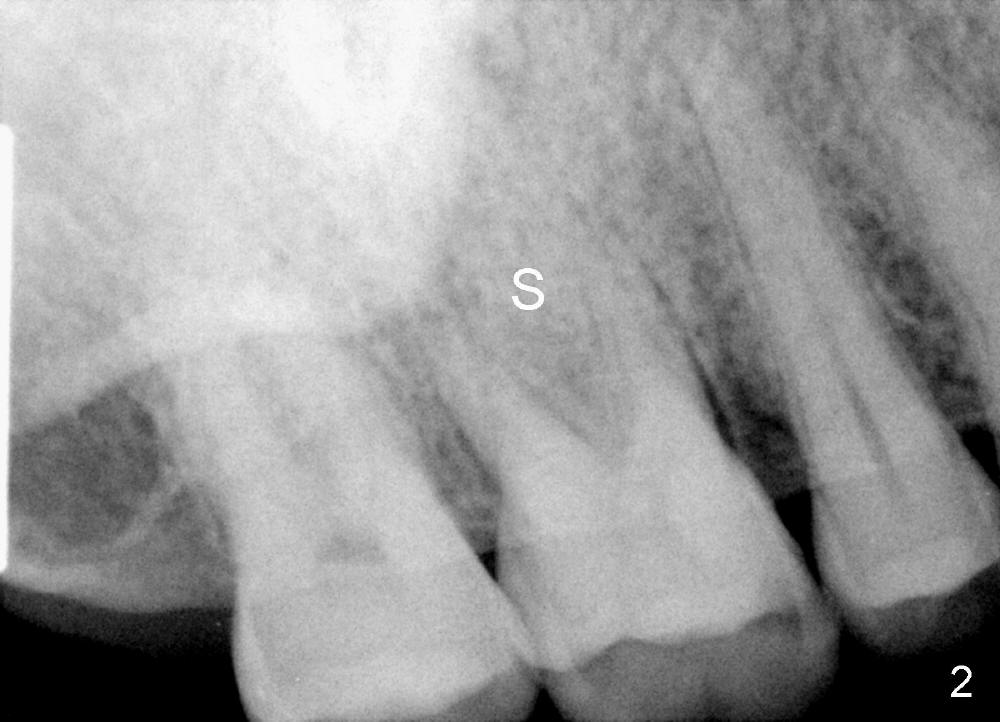
Treatment Plan: Small Immediate Implant Placed in the Septum
A 49-year-old man requests implant restoration of the upper 1st right molar with crack (Fig.1 ^). He loves to chew nuts. There is exosteotosis. In fact the upper left molar also has crown fracture. Bone density should be high. There is abundant bone all around. The limiting factor will be the mesiodistal width of the septum (Fig.2 S), which is 4.87 mm. Bone expansion may be required to place 5 mm implant. The coronal part of the proximal surfaces of the implant may be exposed. Surgical handpiece may be needed to flatten the pointed coronal part of the septum before a 1.5 mm pilot drill is used to initiate ostetomy. Bicon reamers are used to collect bone from the septum of #3. Or osteotomes may be used to expand the osteotomy before implant placement. Then place the autogenous bone with or without allograft back to the three remaining sockets and cover the thread exposed proximal surface. The opening of the sockets will be covered by a collagen membrane, which is fixed between the crest level implant (5.0 mmx14 mm) and healing abutment (8 or 9 mm in diameter). What happens in surgery?
Xin Wei, DDS, PhD, MS 1st edition 01/22/2014, last revision 07/14/2018