
|
|
|
|
|
||||
|
|
|
|
|
|
|||
|
|
|
|
|
|
|||
|
|
|
|
|
|
|
||
Grafting Following Immediate Implant (Photos Provided by Xue Steven, DDS)
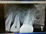
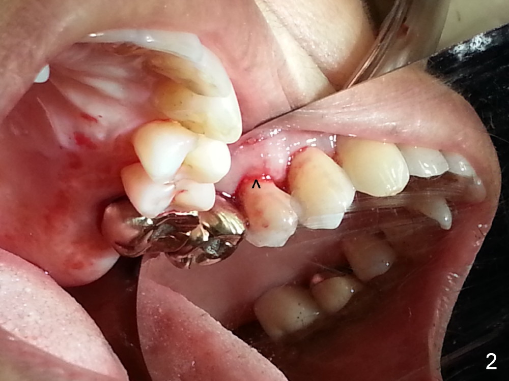
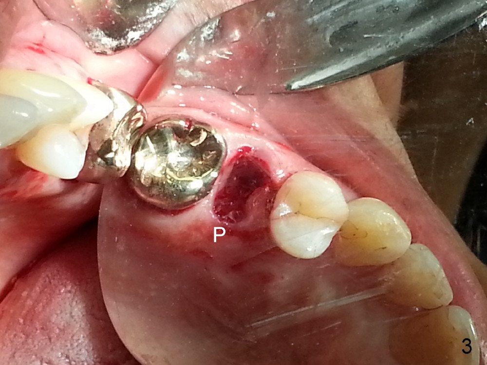
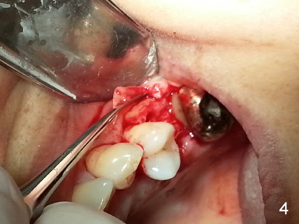
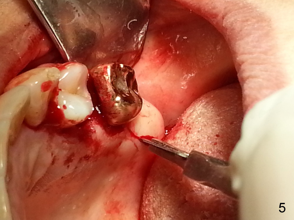
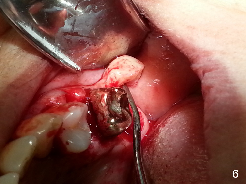
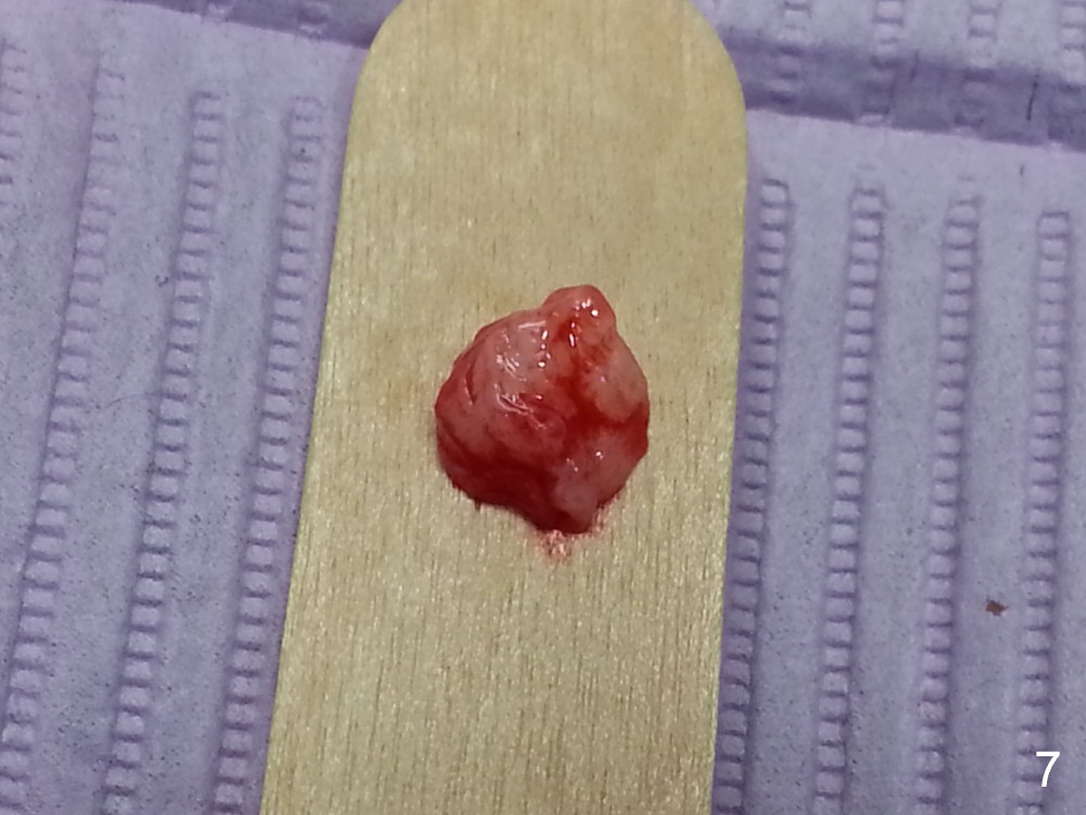
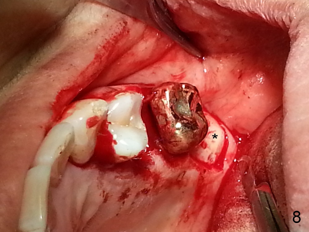
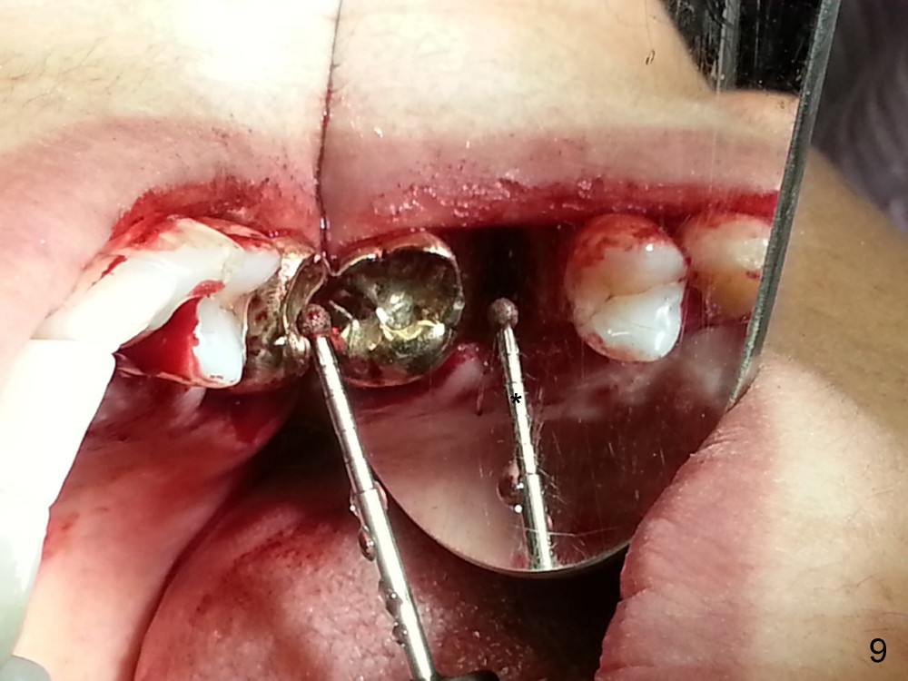
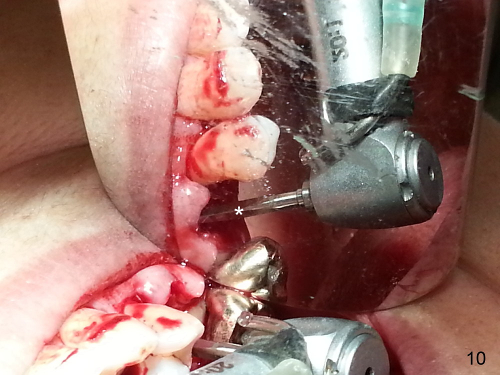
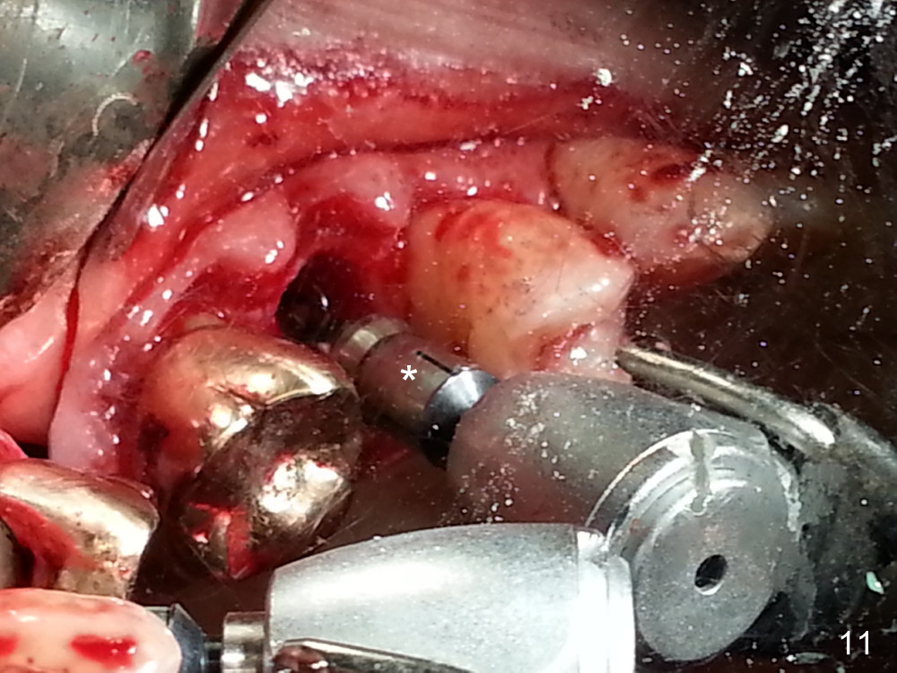
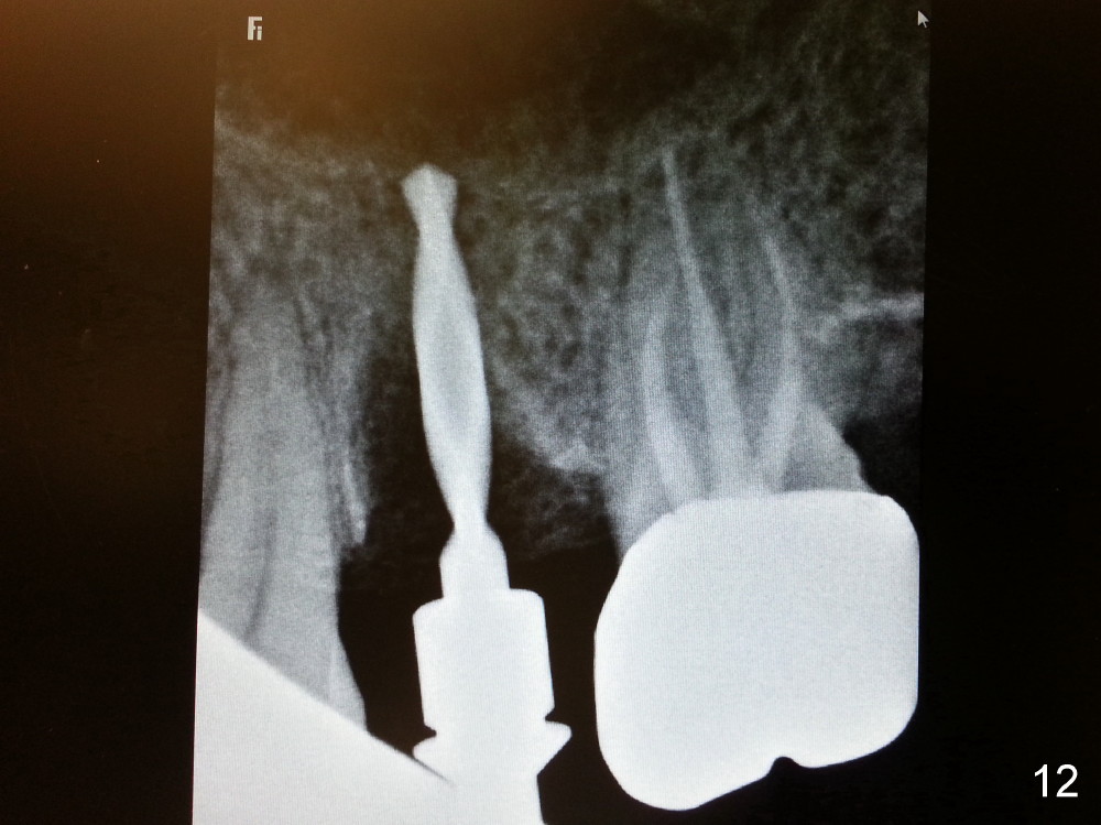
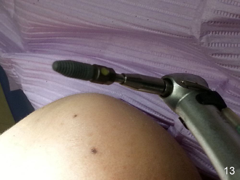
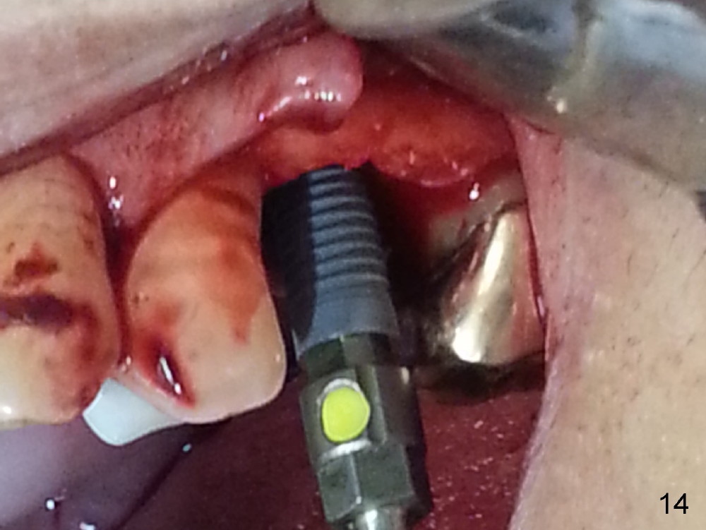
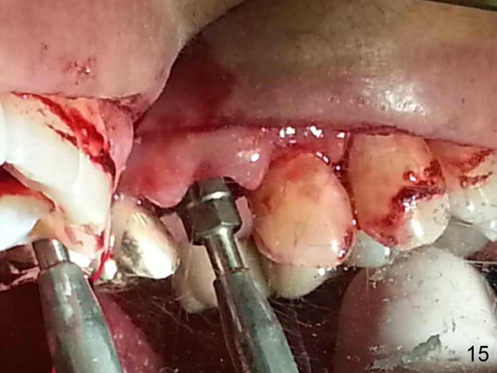
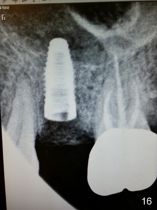
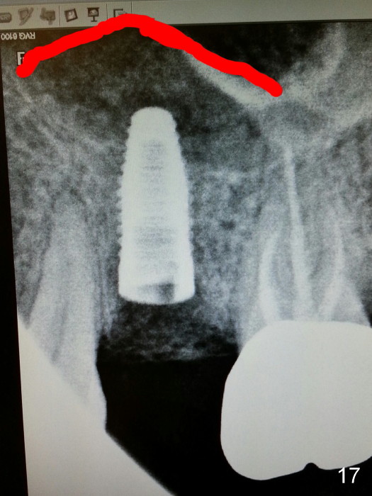
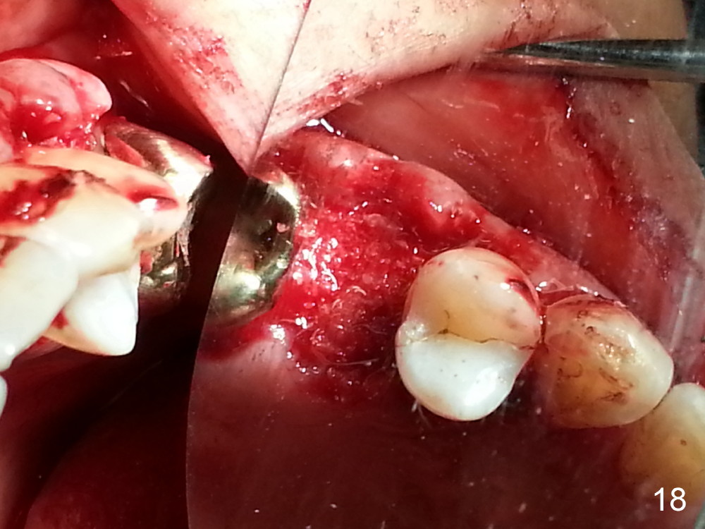
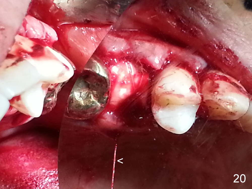
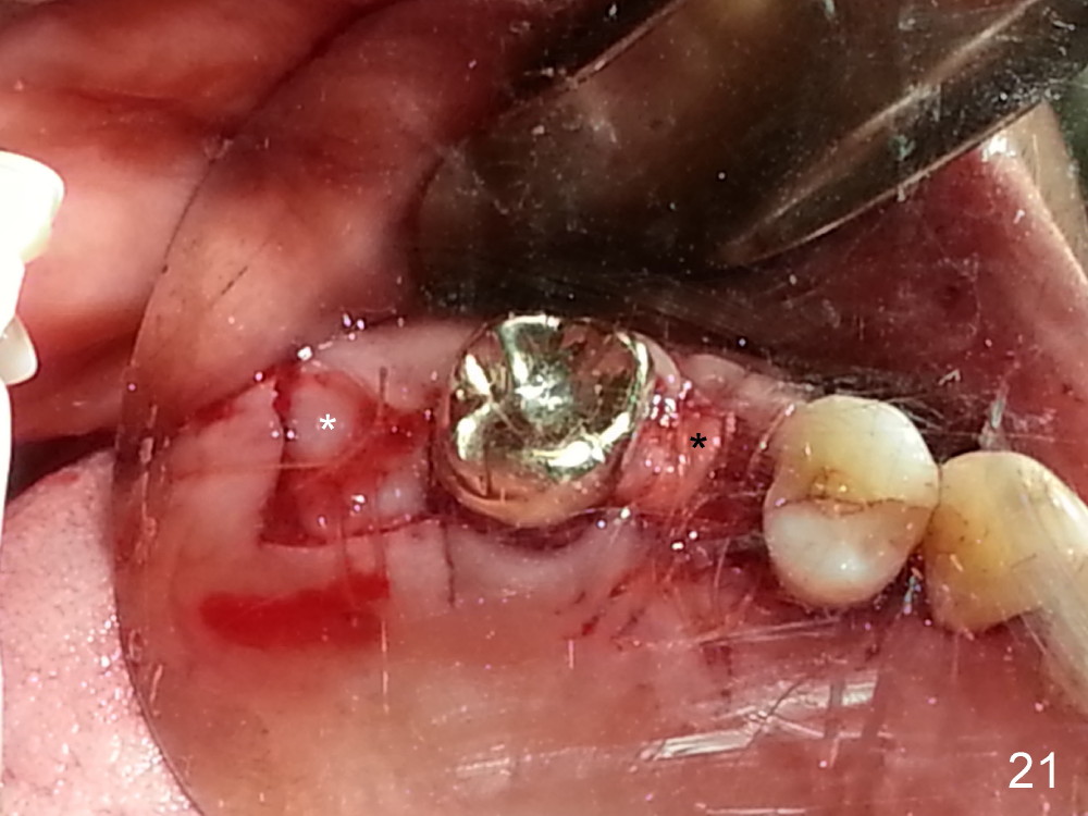
The tooth #13 appears to be affected by periodontits and occlusal trauma (Fig.1*). After using a periotome (Fig.2), the tooth is extracted (Fig.3). The buccal flap is raised (Fig.4). Gingival graft is to be harvested from the site of #15 (Fig.5). The tissue is elevated buccally (Fig.6) and separated (Fig.7). The donor site is covered by a collagen membrane (Fig.8*). A diamond bur is used to induce bleeding from the socket (Fig.9*). Osteotomy is initiated (Fig.10) and enlarged (Fig.11,12). A tapered implant is being placed (Fig.13-15) following internal sinus lift (Fig.16,17). The implant is placed subcrestally, followed by bone graft (Fig.18), soft tissue graft (Fig.19), and suturing (Fig.20 <, Fig.21).
Upper Molar,
Premolar
Immediate Implants
Xin Wei, DDS, PhD, MS 1st edition 02/16/2014, last revision 01/19/2018