|
|
|
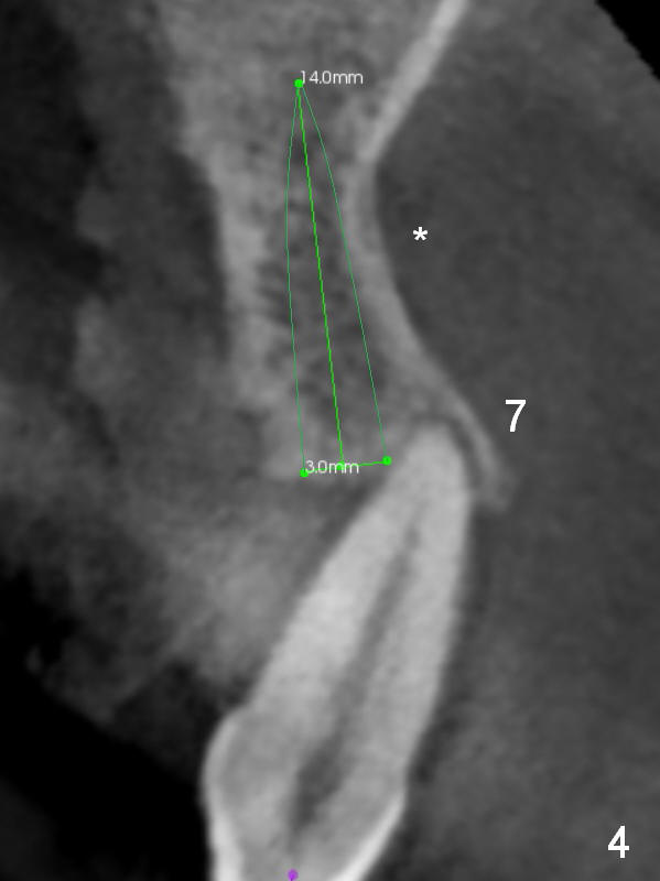
Clinically the tooth #7 is unstable with severe loss of the buccal and palate bone (Fig.4 CBCT coronal section). The tooth will be extracted in the second stage. Note the buccal concavity (*). Palpate the concavity while osteotomy is being made and an implant is being placed. A small implant is going to be placed (3x14 mm).
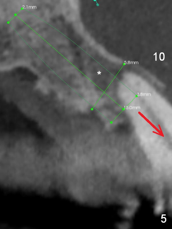
With severe loss of the buccal and palatal bone, the tooth #10 (Fig.5) is incisally shifted (arrow). It appears that there is granulation tissue apically (*, to be removed) . A larger implant than that at #7 will be used. Prepare an angled abutment.
Xin Wei, DDS, PhD, MS 1st edition 02/18/2017, last revision 02/18/2017