|
|
|
|
|
|
|
|
|
|
Bone Expansion
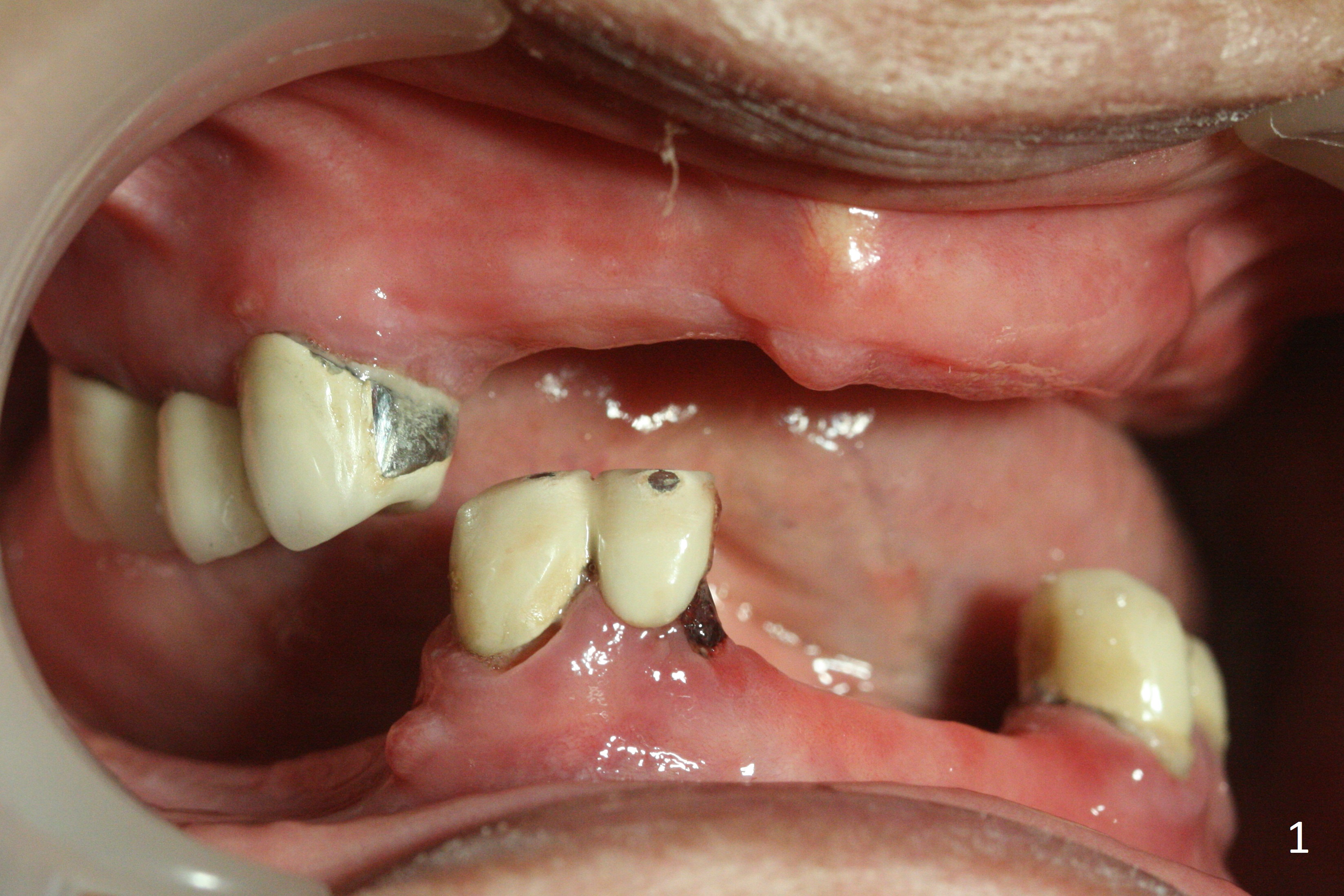
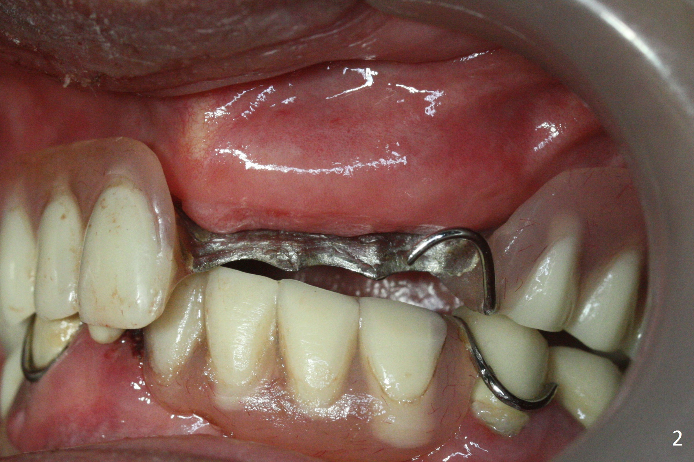
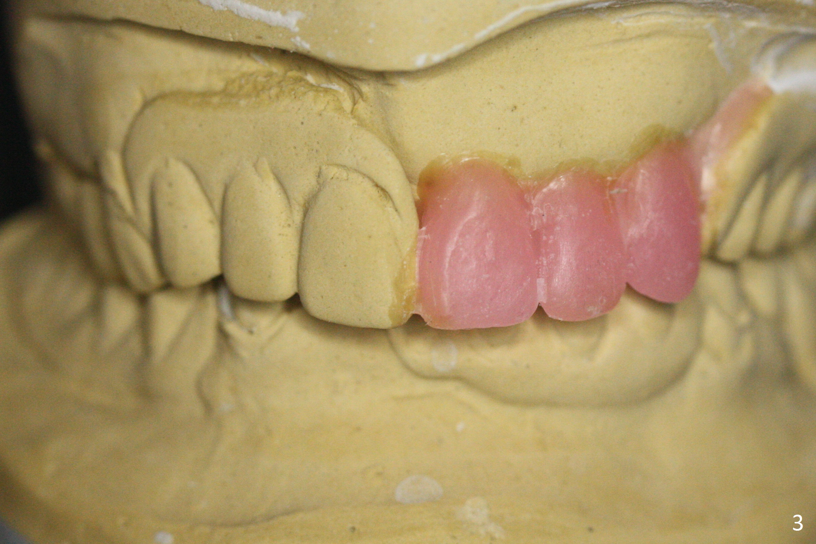
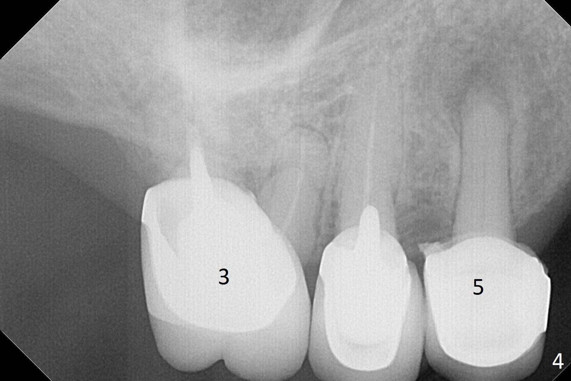
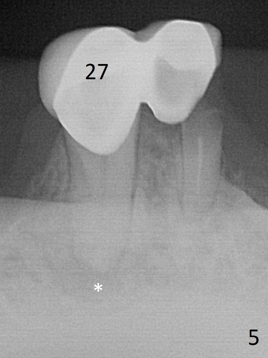
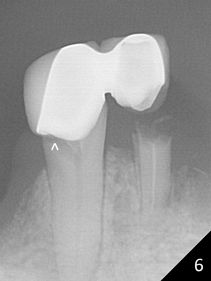
A 62-year-old woman has poor dentition (Fig.1). The most critical area is the upper left anterior: #9-11, which were extracted 4 months earlier (Fig.2). Implants will be placed at #9 and 11 with fabrication of a provisional bridge (Fig.3). After incision, use Magic Split to start bone expansion, followed by micro-osteotomes 1 and 1.5 mm and Magic Osteotomes 3 and 3.8 mm. If the transition between 1.5 and 3 mm osteotomes is not smooth, apply RT2. Place the smallest bone-level implants (for hybrid denture in the future) with gold coated abutments. There should be no interference with or without the partials and in and out. Next step should be removing splinted crowns at #26 and 27 (Fig.1), RCT for #27 and immediate implant at #26 (Fig.5,6 Metronidazole). In fact the splinted crowns at #26 and 27 were lost last week. The patient thinks that she needs 2 implants. The next one is to remove the crown at #5 for RCT (Fig.4).
Return to Upper Arch, Lower Incisor, Canine Immediate Implant, IBS Xin Wei, DDS, PhD, MS 1st edition 03/25/2017, last revision 08/05/2018