.jpg)
|
|
|
|
|
|
|
|
||
Allograft Fills in Defect
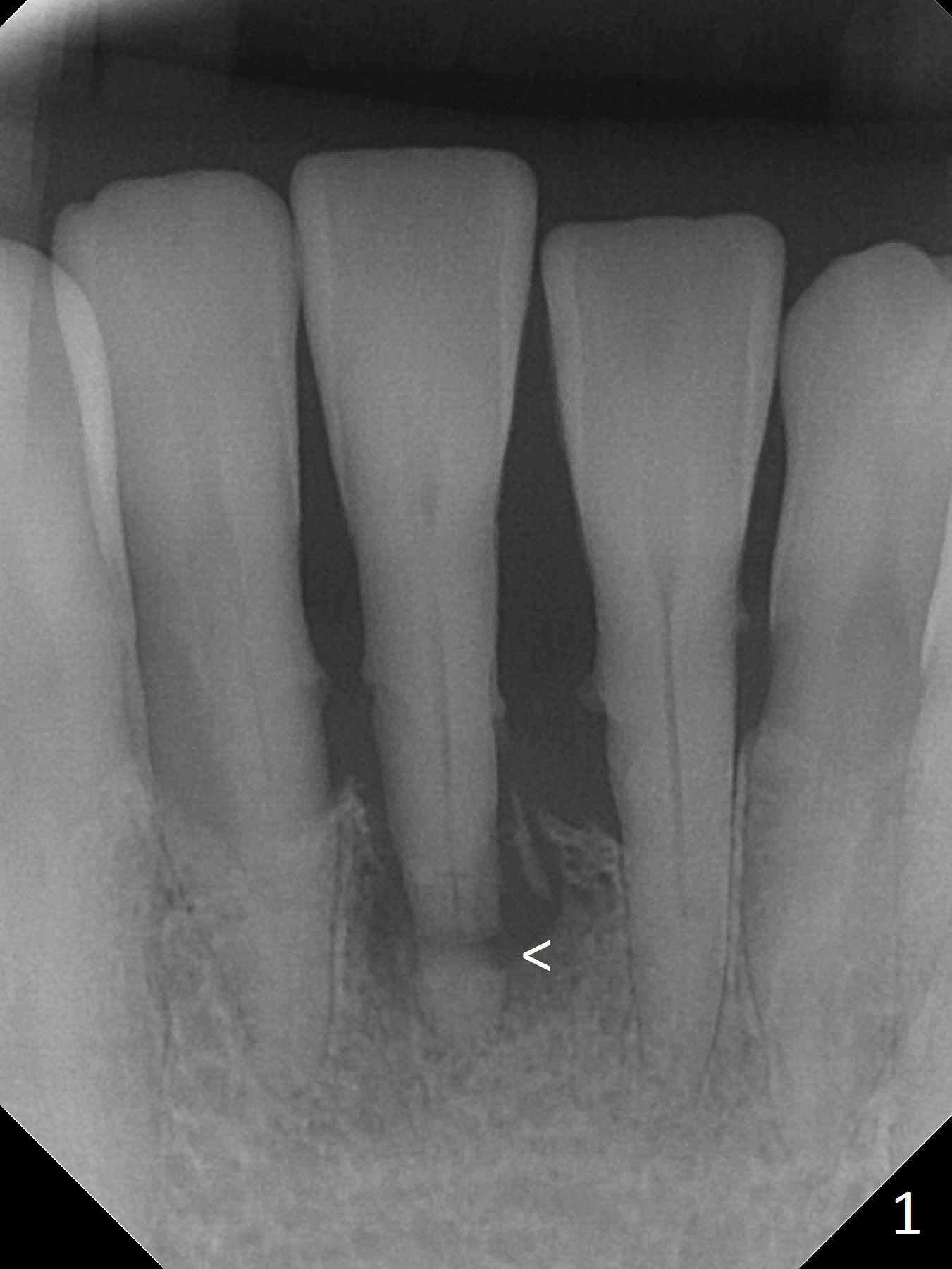
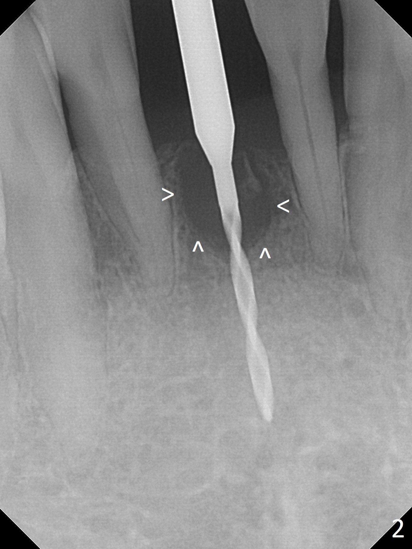
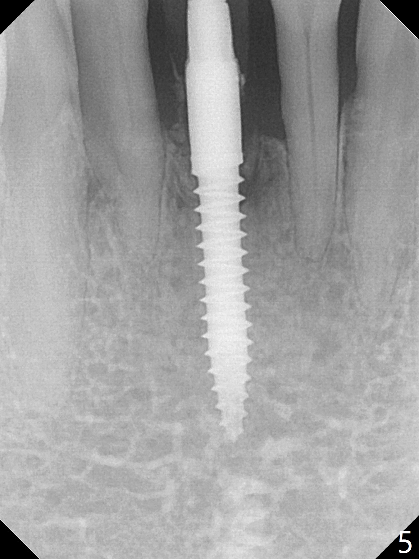
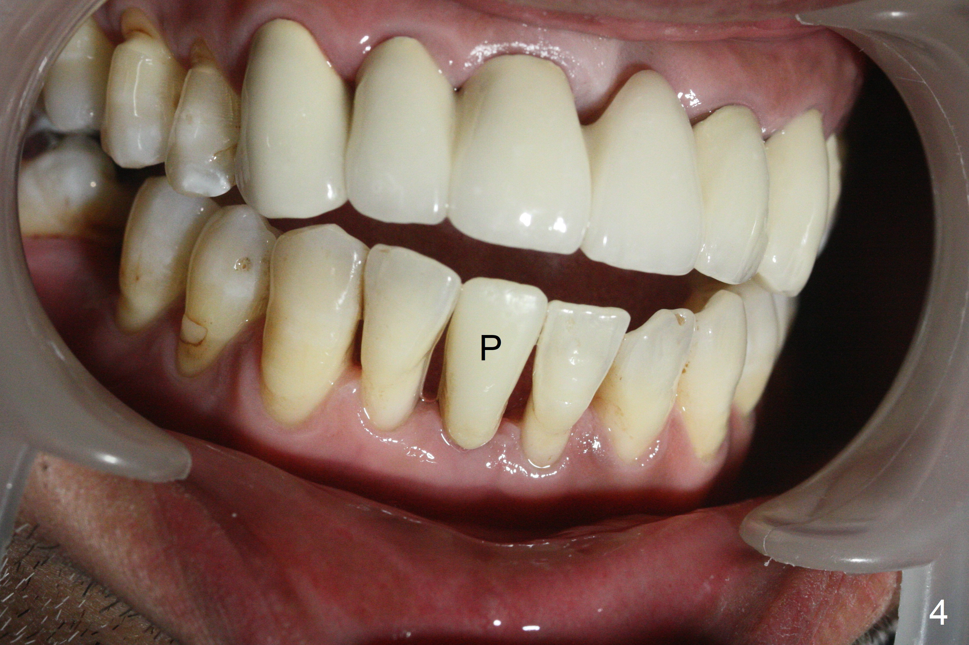
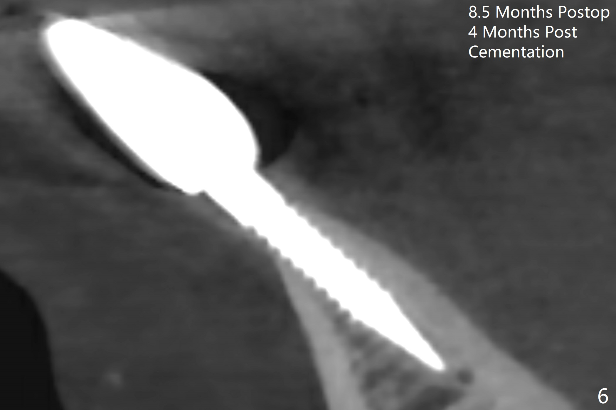
Pre-operative PA shows root tip fracture of the tooth #25 (Fig.1 <). After extraction, the defect is evident (Fig.2 arrowheads). Clinically there is no lingual plate, whereas the buccal one is low. Allograft fills in the defect (Fig.3 *) following placement of a 3x16(4) mm 1-piece implant with insertion torque >50 Ncm. Later the implant is placed deeper to reduce possibility of periimplantitis. With scaling & root planing, the gingiva around the provisional is healthy 3 weeks postop (Fig.4 P). The bone graft persists in the socket 4 months postop (Fig.5). Implant threads seem to be covered by the bone, as revealed by CT taken 8.5 months postop; 4 months post cementation (Fig.6).
Return to
Lower
Incisor Immediate Implant, IBS,
#14
Xin Wei, DDS, PhD, MS 1st edition 06/16/2017, last revision 04/08/2018