|
|
|
|
|
|
|
|
|
|
|
|
Delayed Implant Associated with Tilting Neighboring Teeth M
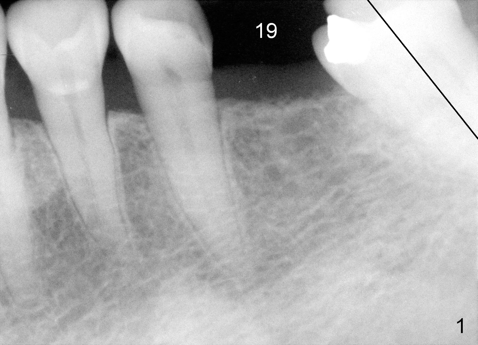
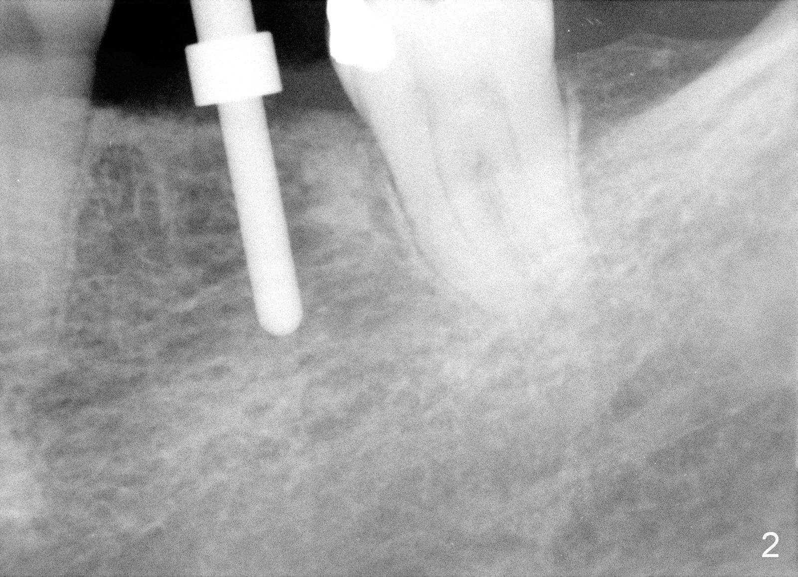
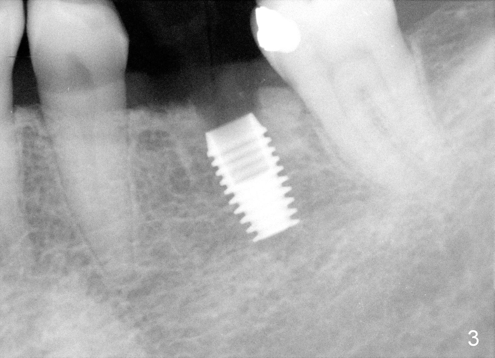
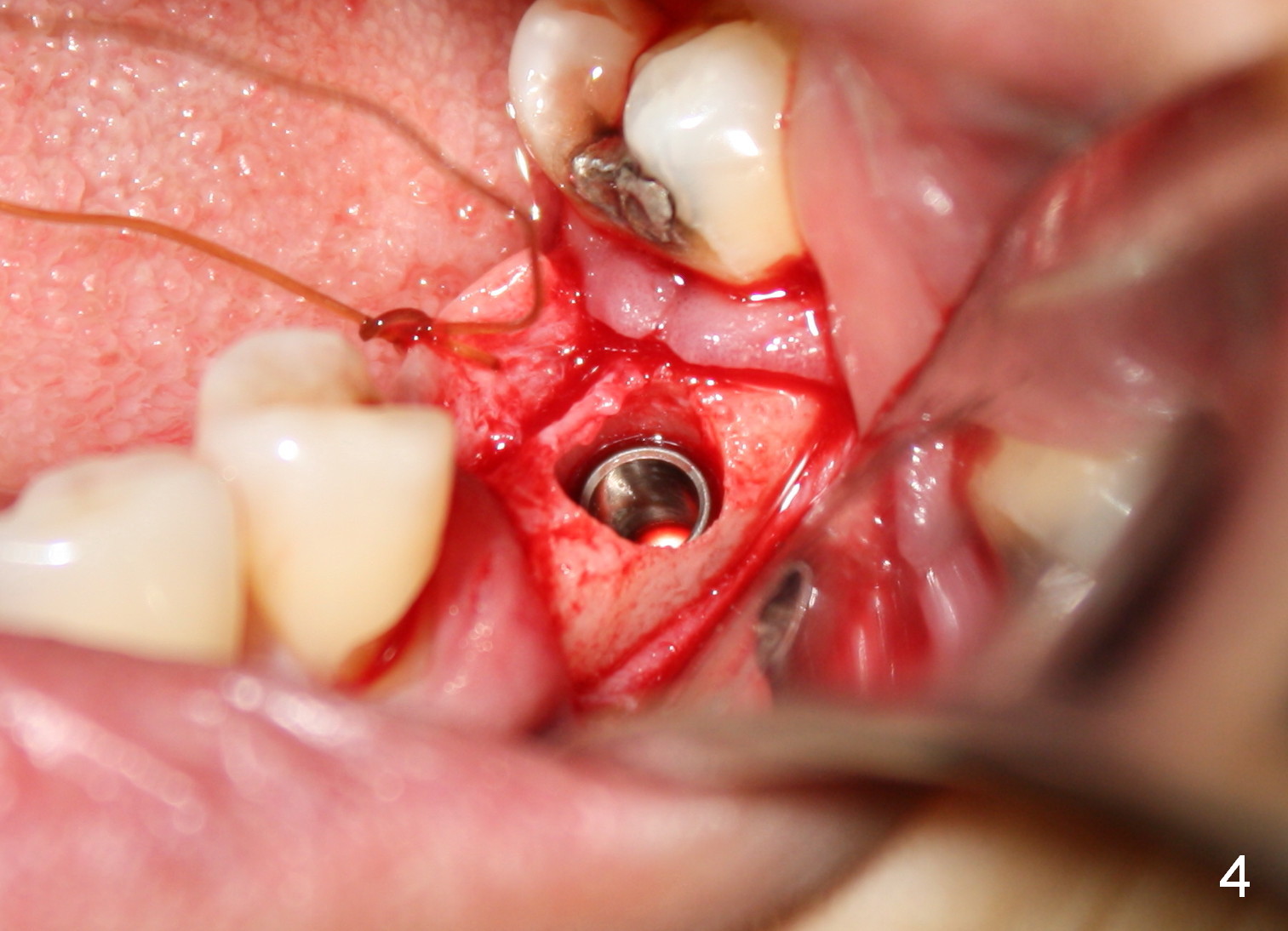
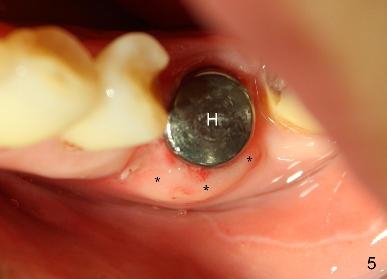
A 68-year-old lady was interested in using an implant at the site of #19 as an anchorage to upright and distalize the 2nd molar (Fig.1 oblique line representing the long axis of the tooth). The implant is intended to be placed distal of the edentulous space (Fig.2). Bicon implant (4.5x8 mm; Fig.3) is chosen because of easy change of abutments as the space increases orthodontically. The ridge is narrow (Fig.4). After packing autogenous bone and placing collagen membrane coronal to the implant, a healing abutment is placed immediately (5x6.5mm); by 15 days postop, a gingival cuff has formed (Fig.5 *) buccal to the healing abutment (H).
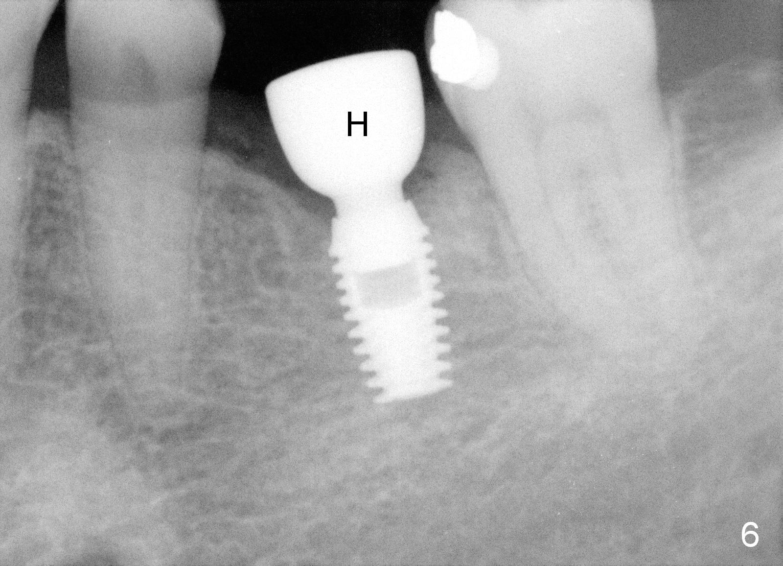
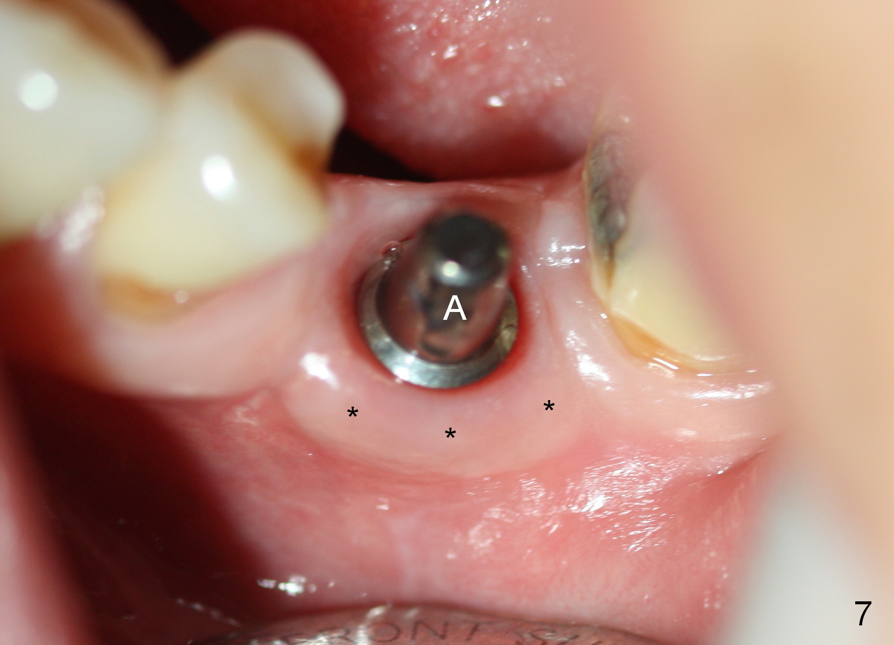
The implant appears to osteointegrate 3.5 months postop (Fig.6). When a restorative abutment is installed (Fig.7 A), the gingival cuff (*) remains distinct.
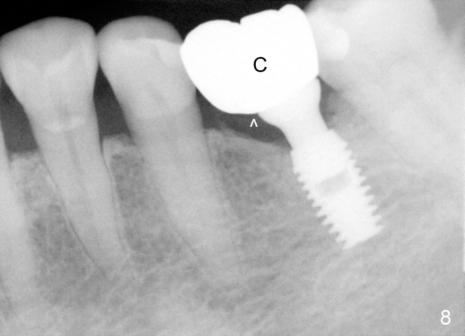
Suddenly her schedule becomes busy. She is not so eager to have limited ortho. A definitive restoration is placed (Fig.8 C). Bicon crown can be cemented intra- or extra-orally. The former and traditional way is used for this case, since the margin is equi- or slightly sub-gingival (Fig.7). After cementation, a PA is taken to confirm no residual cement (Fig.8 ^). Otherwise, the crown/abutment complex could be removed for cement removal and reseated.
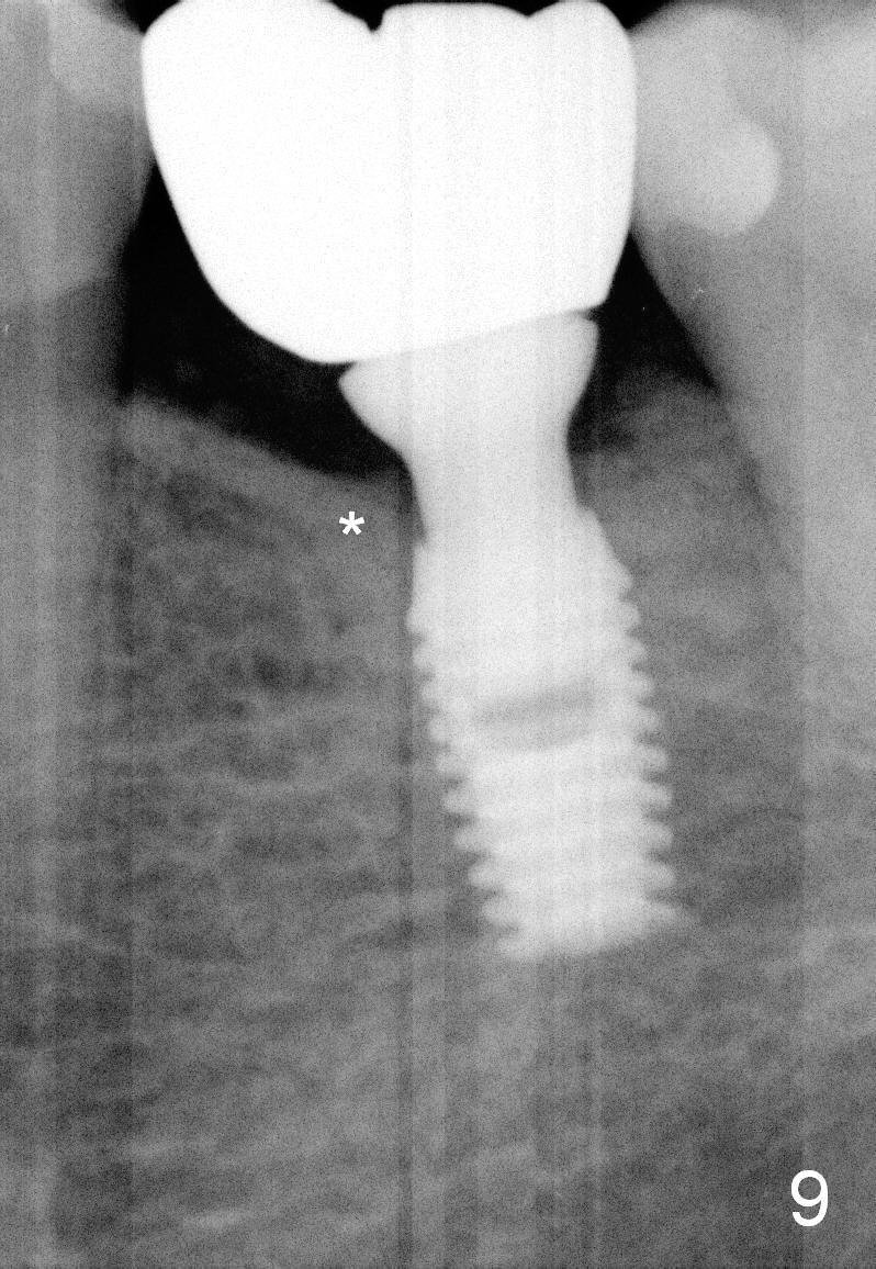
One year post cementation, the mesial bone appears to be higher than before (Fig.9 *).
Return to Lower Molar Immediate Implant,
Dr.
Wu, Thread
Patterns,
Implant & Ortho
2
#15
IBS Intraop Bone Graft
Xin Wei, DDS, PhD, MS 1st edition 04/19/2015, last revision 01/01/2020