


|
|
|
|
|
|
|
|
|
|
||
|
|
|
|
|
|
Ridge Split with Wheel Saw
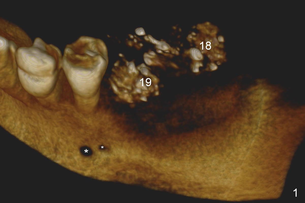
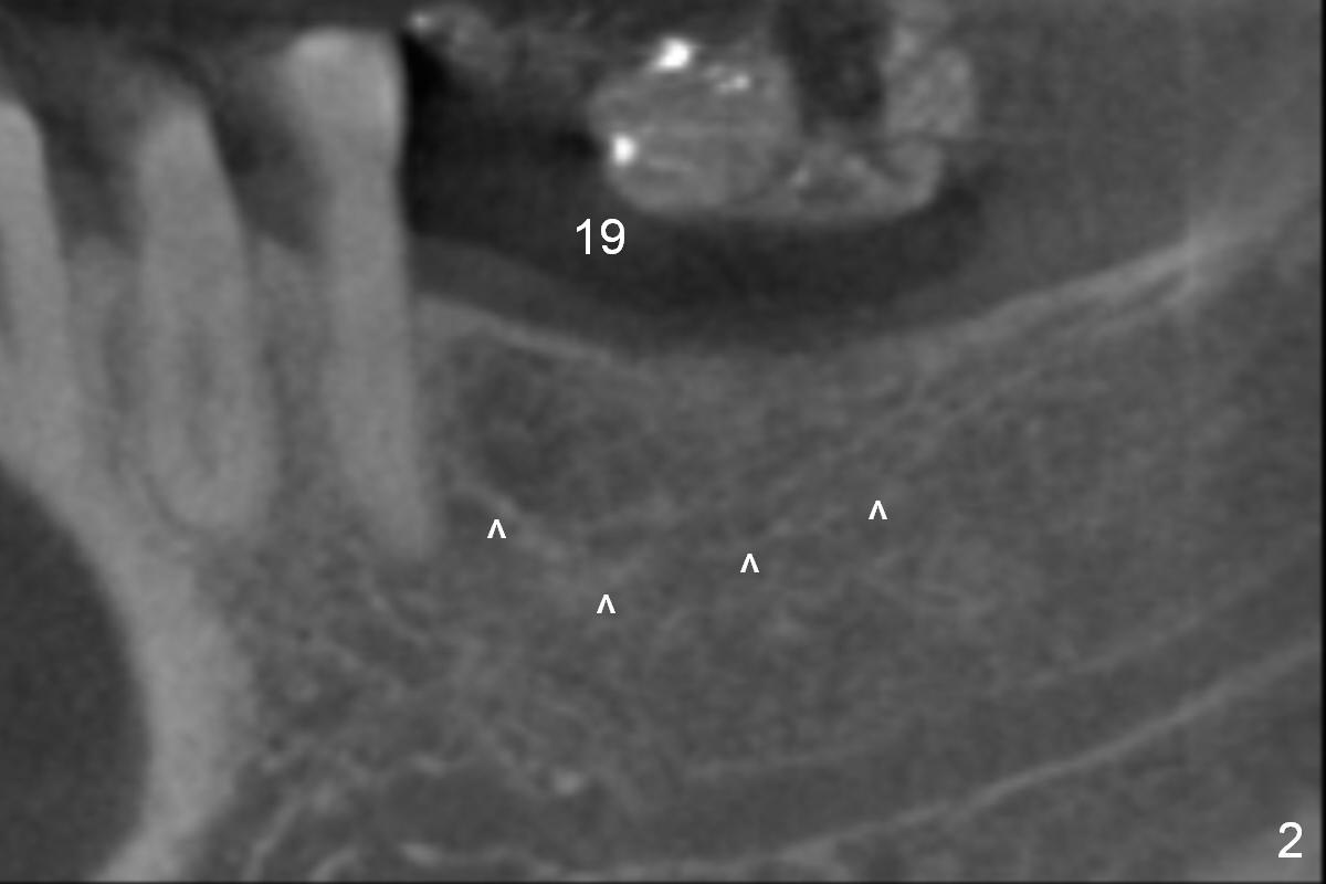
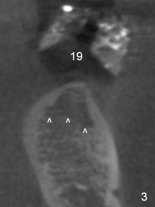
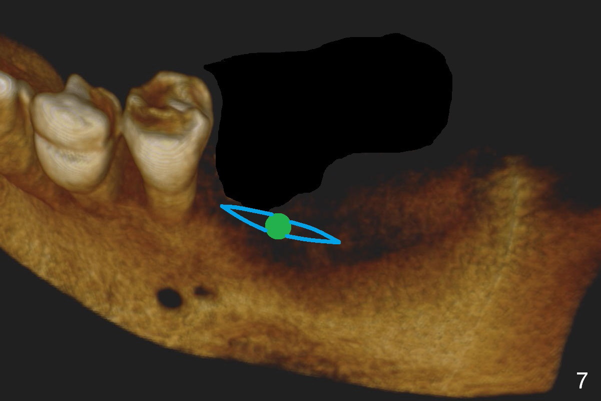
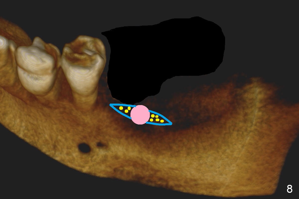
CBCT 3-D image taken with scan stents at the sites of #18 and 19 shows 2 Mental Foramina (Fig.1 *) in the left mandible. Both sagittal (Fig.2) and coronal (Fig.3) sections show low density at the crest of #19 of a 55-year-old man (arrowheads). The crest is also narrow. After making an initial osteotomy through the stent (Fig.4 red arrow), remove the latter (Fig.5). Ridge split is initiated with wheel saws (Fig.6 blue line). The osteotomy is enlarged with bone expanders (Fig.7 green circle) with ridge split (blue lines). Finally an implant is placed (Fig.8 pink circle) with placement of bone graft in the space of the ridge split (yellow circles).
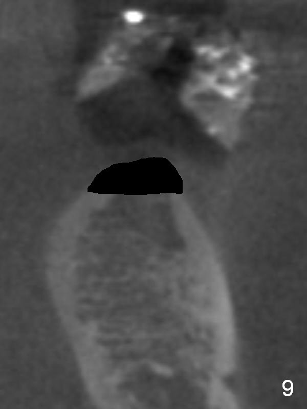
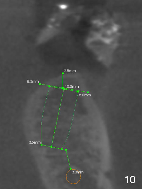
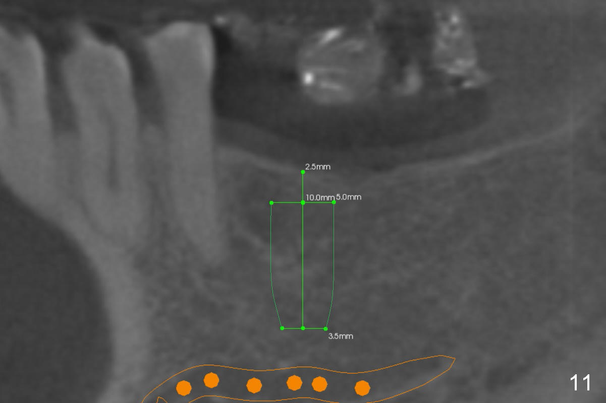
Without ridge split, the ridge top has to be trimmed (Fig.9 black area) to hold a 3 mm shorter implant (Fig.10,11).
Return to Lower Molar Immediate Implant
Xin Wei, DDS, PhD, MS 1st edition 03/27/2016, last revision 03/27/2016