,%20bone%20graft,%20Osteotape.jpg)
|
|
|
|
|
|
|
|
|
|
||
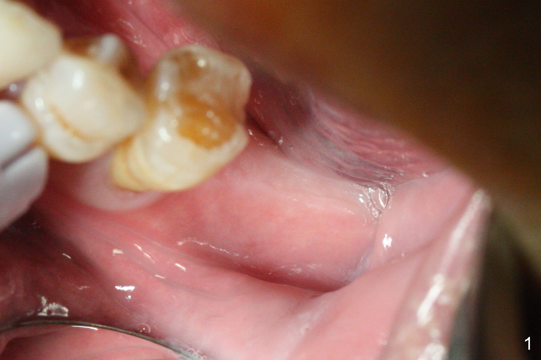
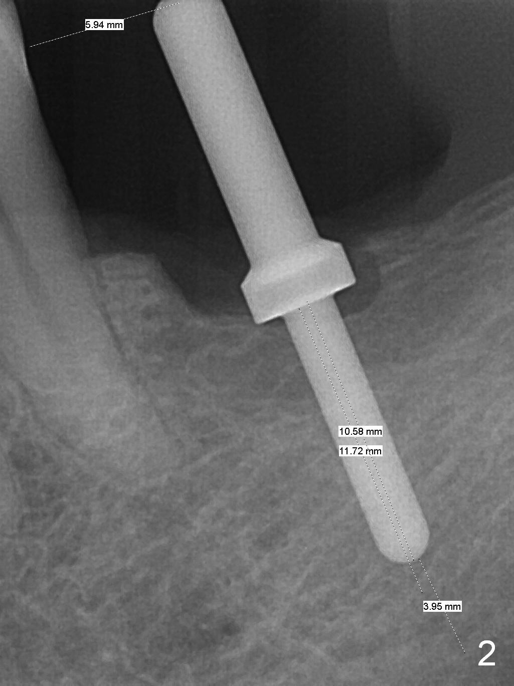
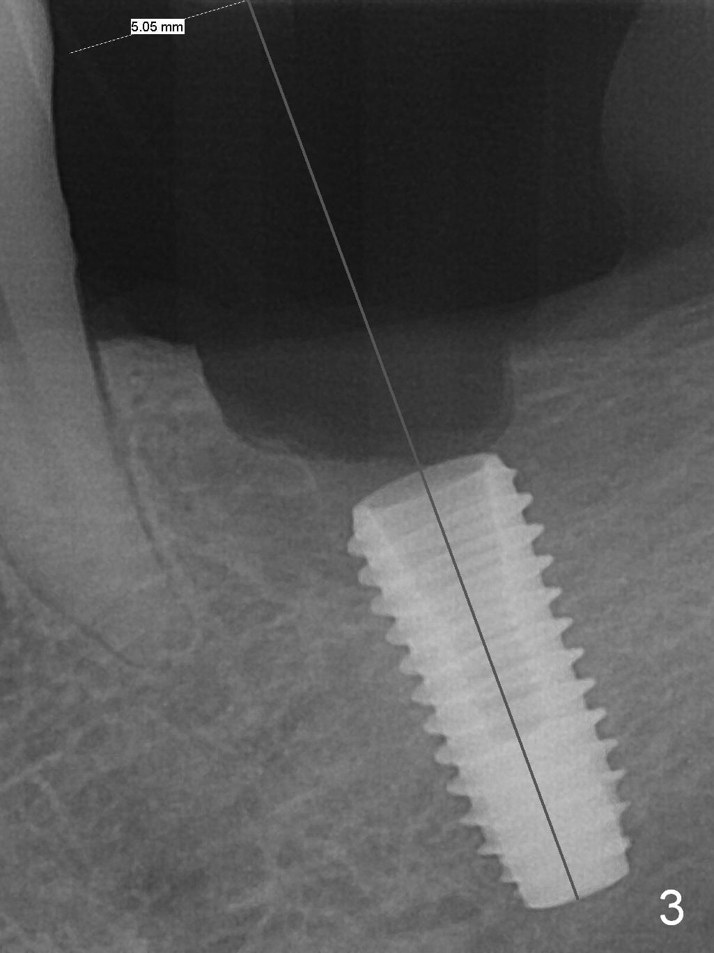
Simultaneous Implant Placement and GBR
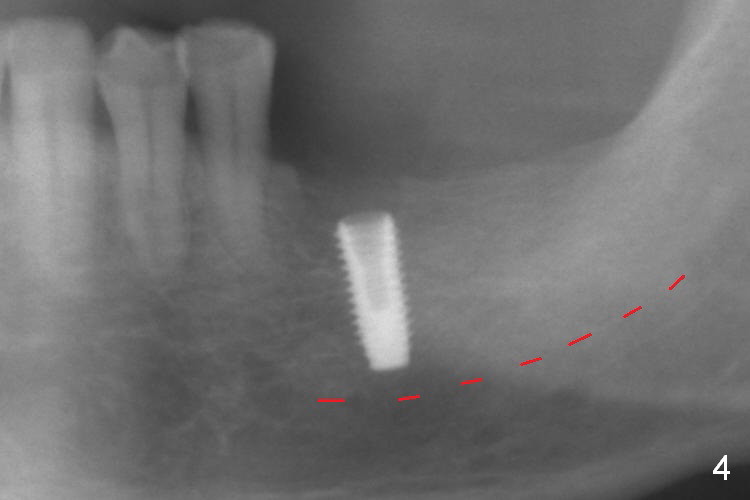
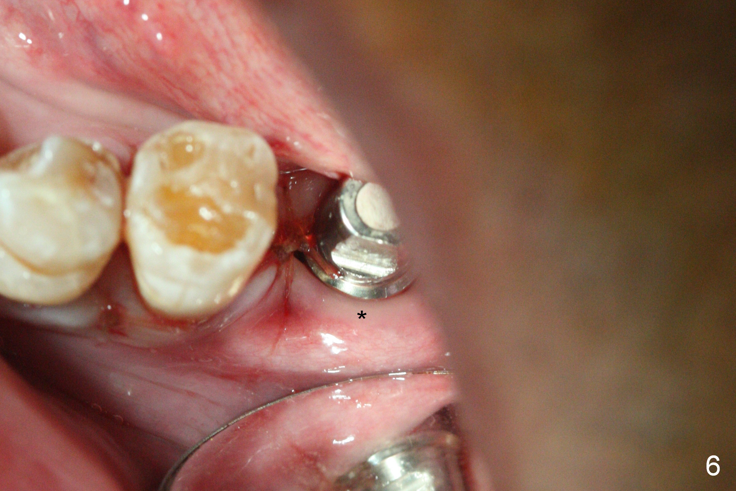
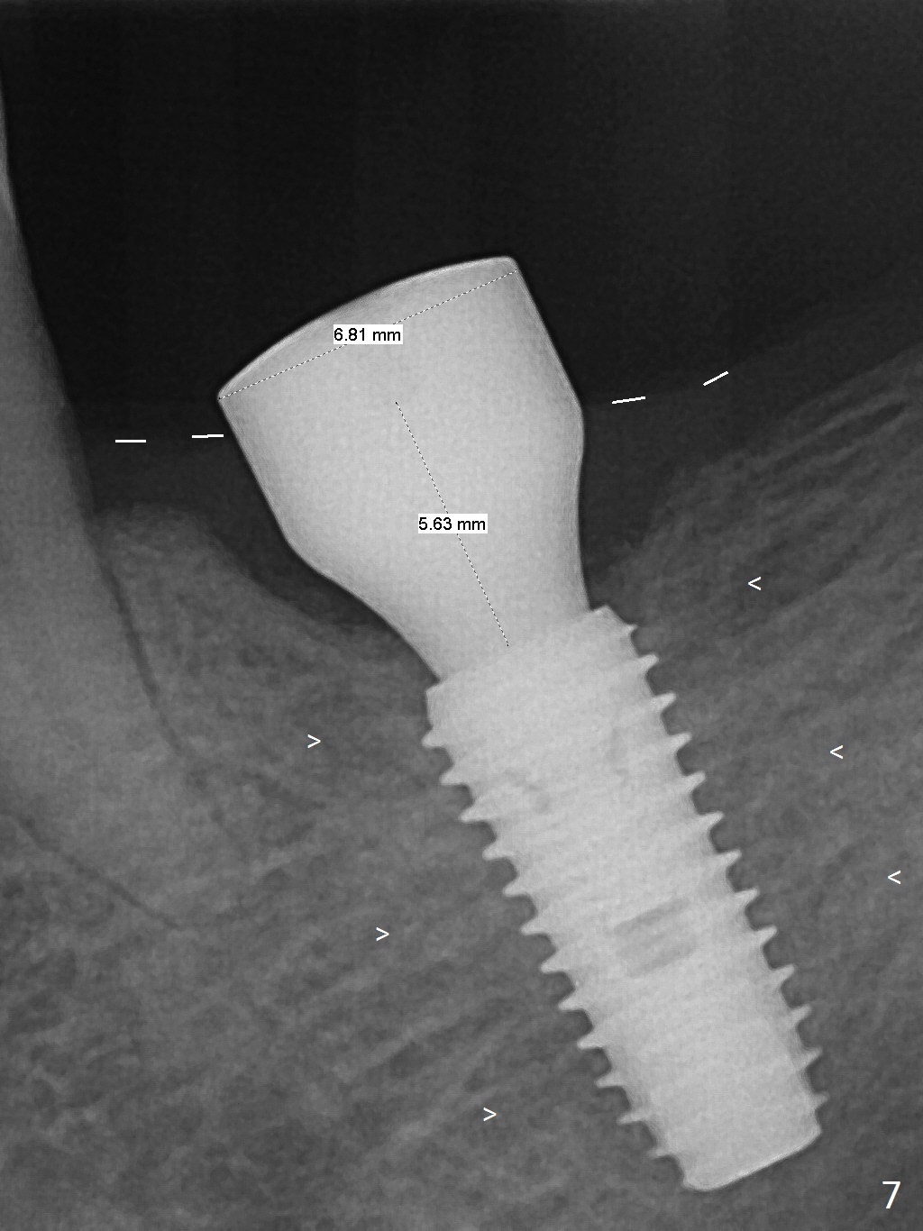
As indicated by CBCT, the ridge at the site of #19 is pointed (Fig.1). After regional ridge reduction (Fig.2) and use of 2 mm pilot drill for 8.5 mm, the depth is actually ~ 10 mm. After sequential osteotomy until 4.3x10 mm drill, the osteotomy is in fact 11.5 mm deep; a 5x11.5 mm implant is placed with <35 Ncm (Fig.3). Panoramic X-ray shows the implant close to the superior border of the Inferior Alveolar Canal (Fig.4 red dashed line). The ridge looks wider and more bulging (*) when a 6.5x5.5(5) mm abutment, allograft/Osteogen and Osteotape (GBR) are placed (Fig.5 (lingual) and 6 (buccal)). Periodontal dressing is then applied. There is no postop paresthesia. The abutment dislodges 3 weeks postop. A healing abutment is placed (6.5x4 mm). The patient returns for restoration 4 months postop; bone density appears to increase around the implant (Fig.7 arrowheads). When a 7.5x5(4) mm abutment is placed, there is transient pressure on the gingiva (blanching). The mesial gingival trough is formed by Diode laser prior to impression.
Return to
Lower
Molar Immediate Implant, IBS,
#3
Xin Wei, DDS, PhD, MS 1st edition 01/30/2017, last revision 06/04/2017