|
|
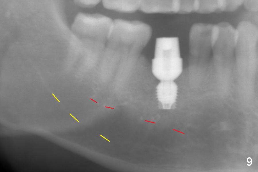
When bone graft (Fig.7 *) and 7.8x5.5(6) mm abutment (Fig.7,8) are placed, panoramic X-ray is taken (Fig.9). There appears to be a thick layer of spongy bone in the posterior mandible between the red and yellow dashed lines.
Xin Wei, DDS, PhD, MS 1st edition 03/14/2017, last revision 06/11/2017