.jpg)
|
|
|
|
|
|
|
|
|
|
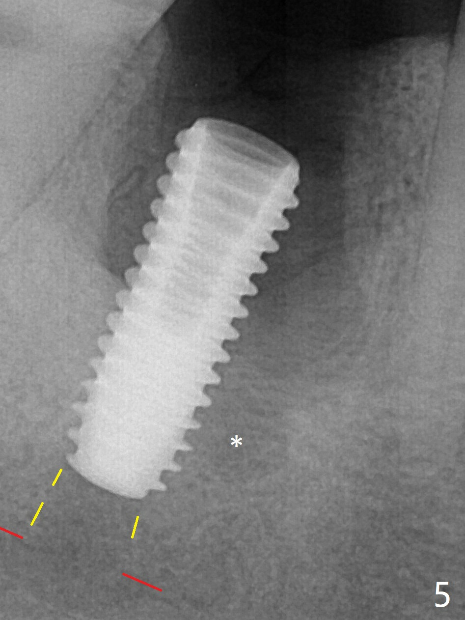
A 5x13 mm implant is placed superficially (Fig.5) with a trace of the previous osteotomy (yellow line) and deep space created by the mesial osteotomy (*). Red dashed line: the superior border of the Inferior Alveolar Canal. Apparently the pathological and iatrogenic defects are filled with allograft (Fig.6 *). Guided surgery could have avoided the mesial osteotomy.
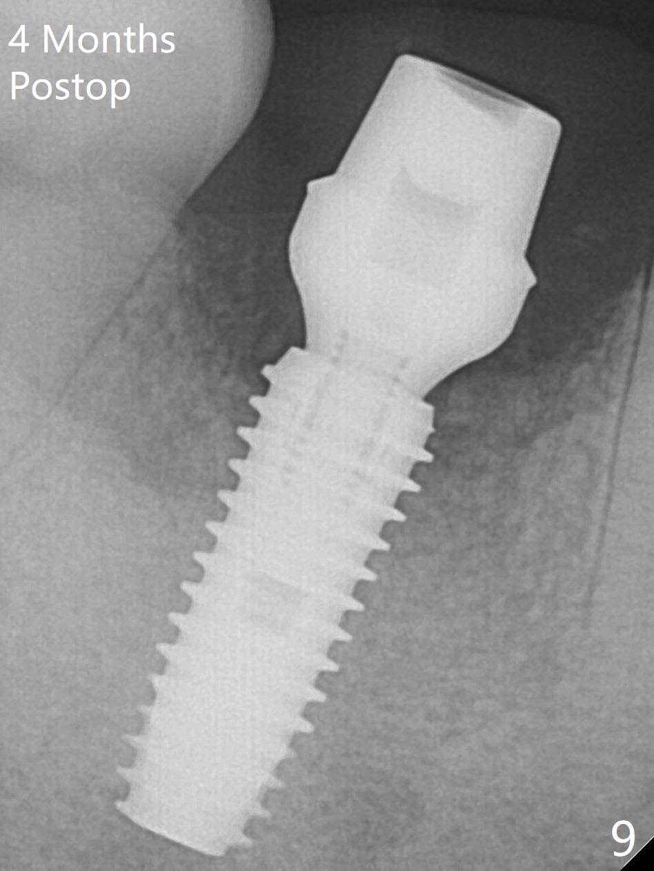
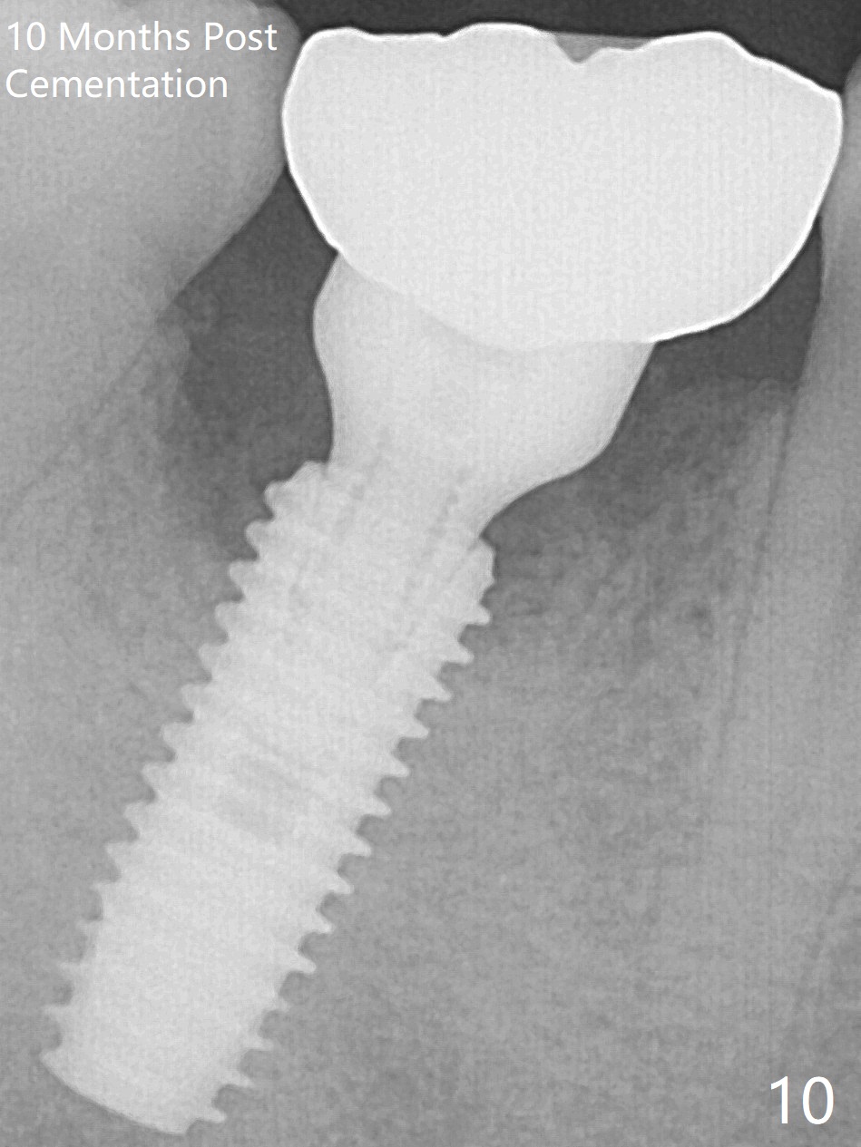
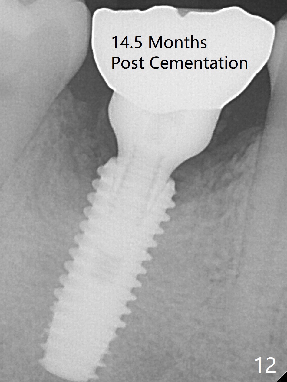
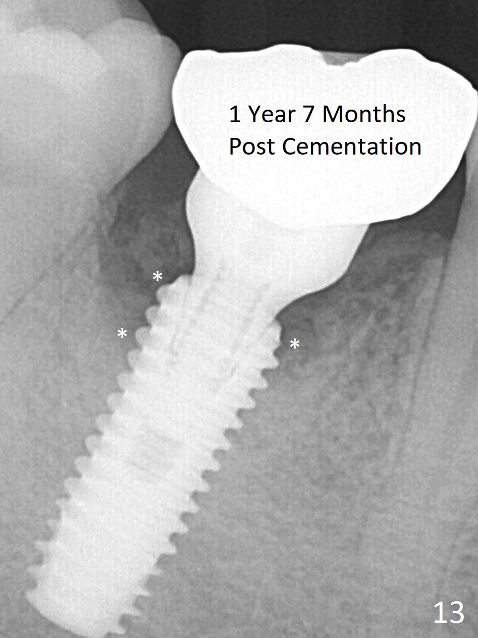
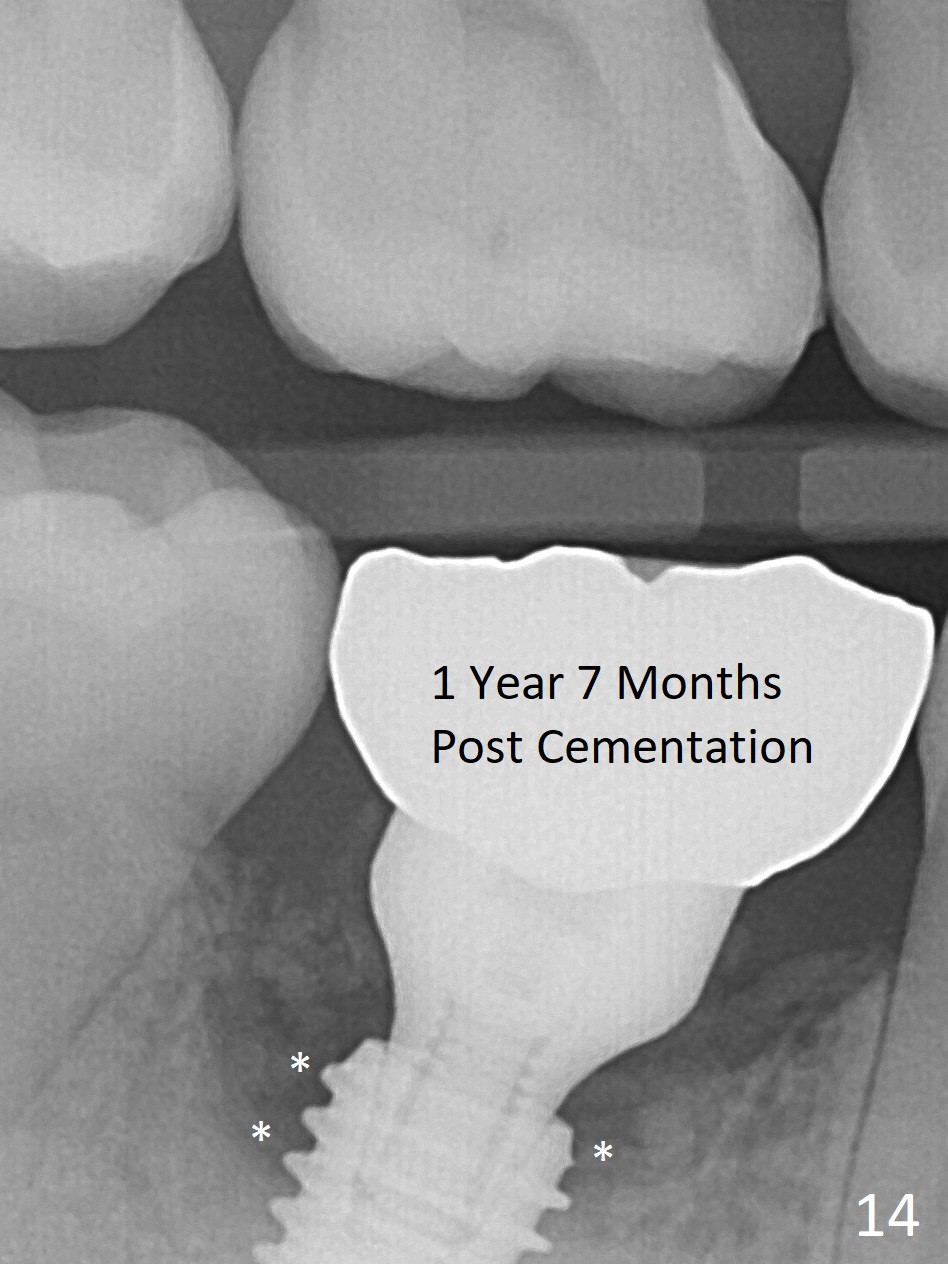
Bone morphology at the coronal end of the implant apparently changes 4 months postop, suggesting osteointegration (Fig.9). Impression is taken after change in abutment from 6.5x4(4) mm to (5) with prep. Bone density around the implant at the crest seems to increase 10 months post cementation (Fig.10). The bone appears to regenerate toward the abutment, particularly distally, 14.5 months post cementation (Fig.12). Periimplantitis develops (bleeding on water pik and erythematous and tender buccal gingiva) 1 year 7 months post cementation (Fig.13,14). The 1st three threads are exposed (*). Bone graft with PRF and 6-month membrane or Cytoplast will be needed.
Long Implant Placed Deep Last Next
Xin Wei, DDS, PhD, MS 1st edition 06/17/2019, last revision 07/13/2019