
|
|
|
|
|
|
|
|
|
|
||
|
|
|
|
|
|
|
|
|
|
||
|
|
|
|
|
|
|
|
|
|
||
|
|
|
|
||
|
|
||||
Single Drill Stabilizes Osteotomy
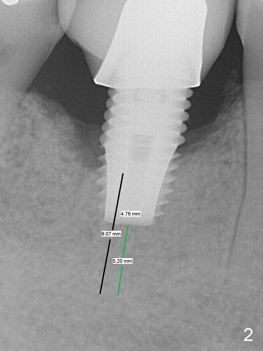
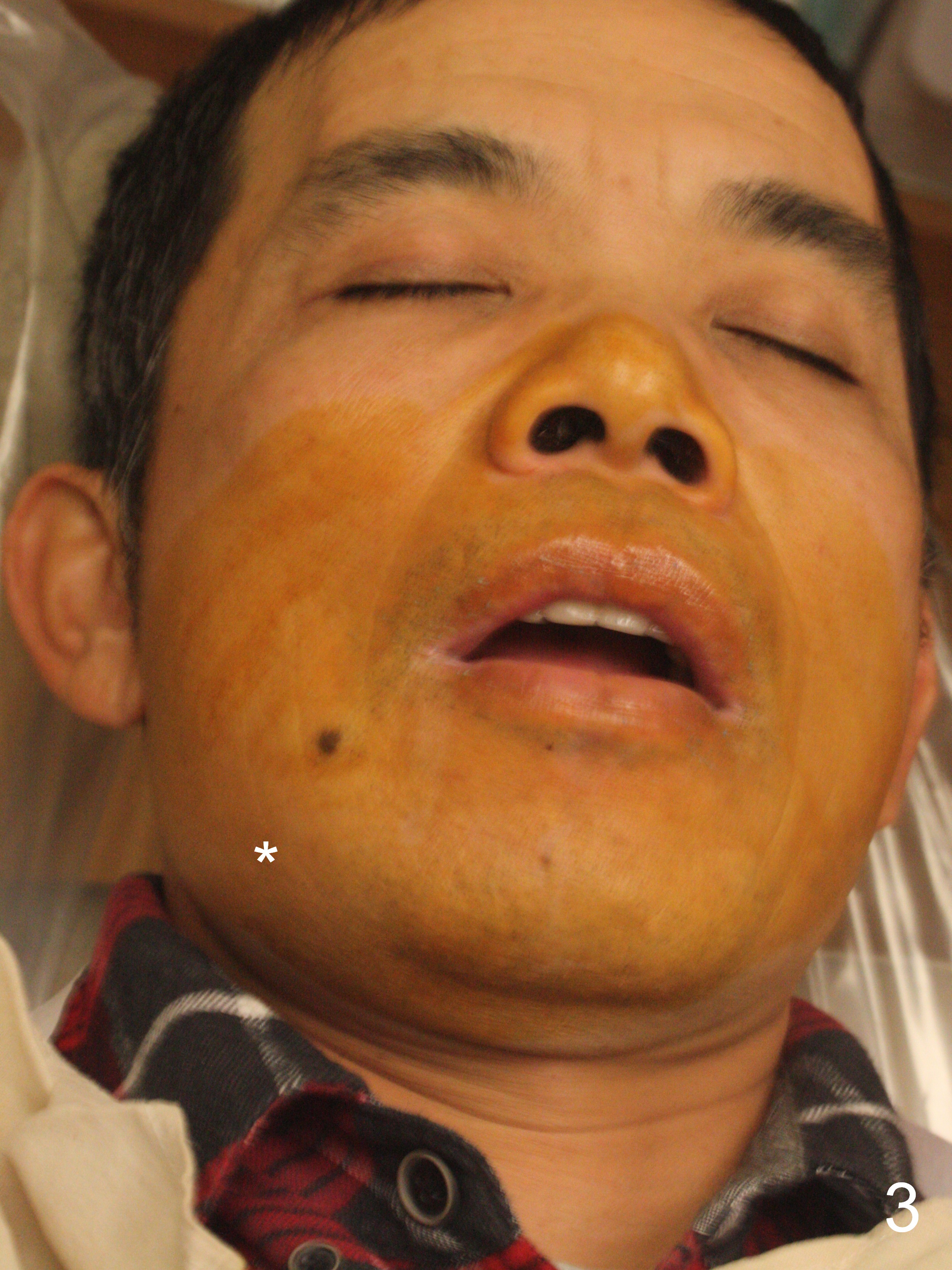
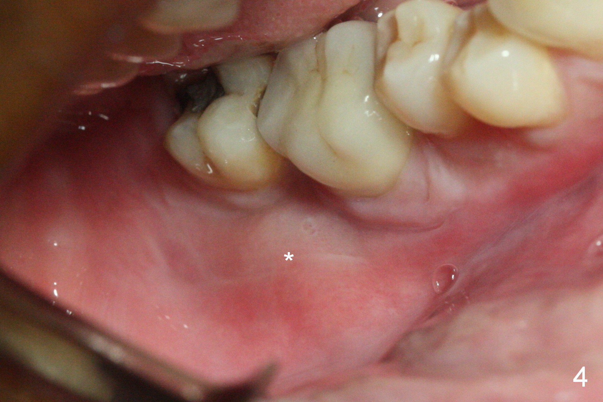
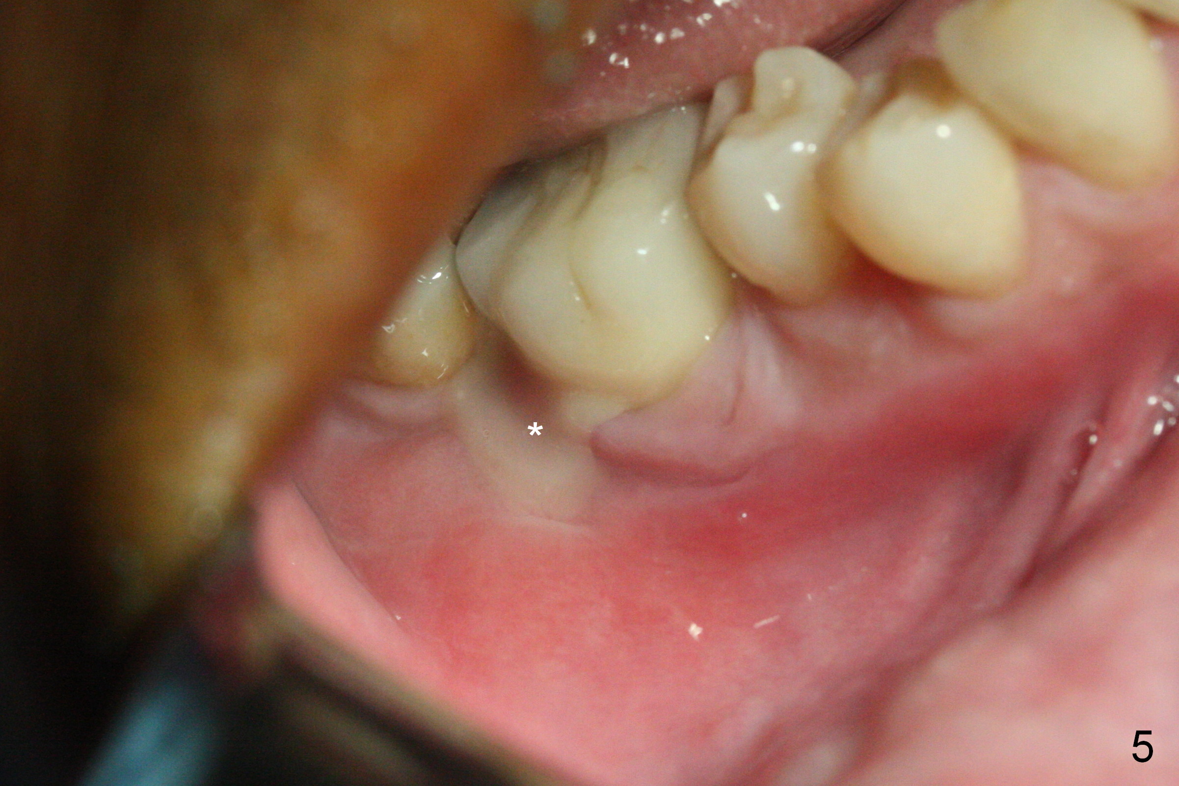
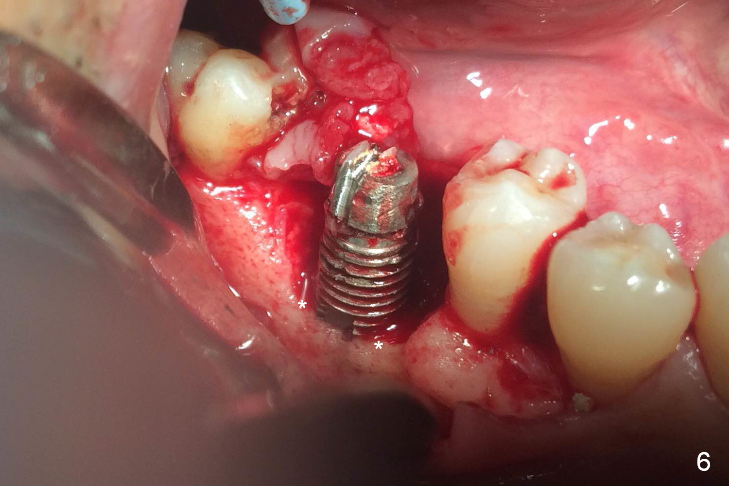
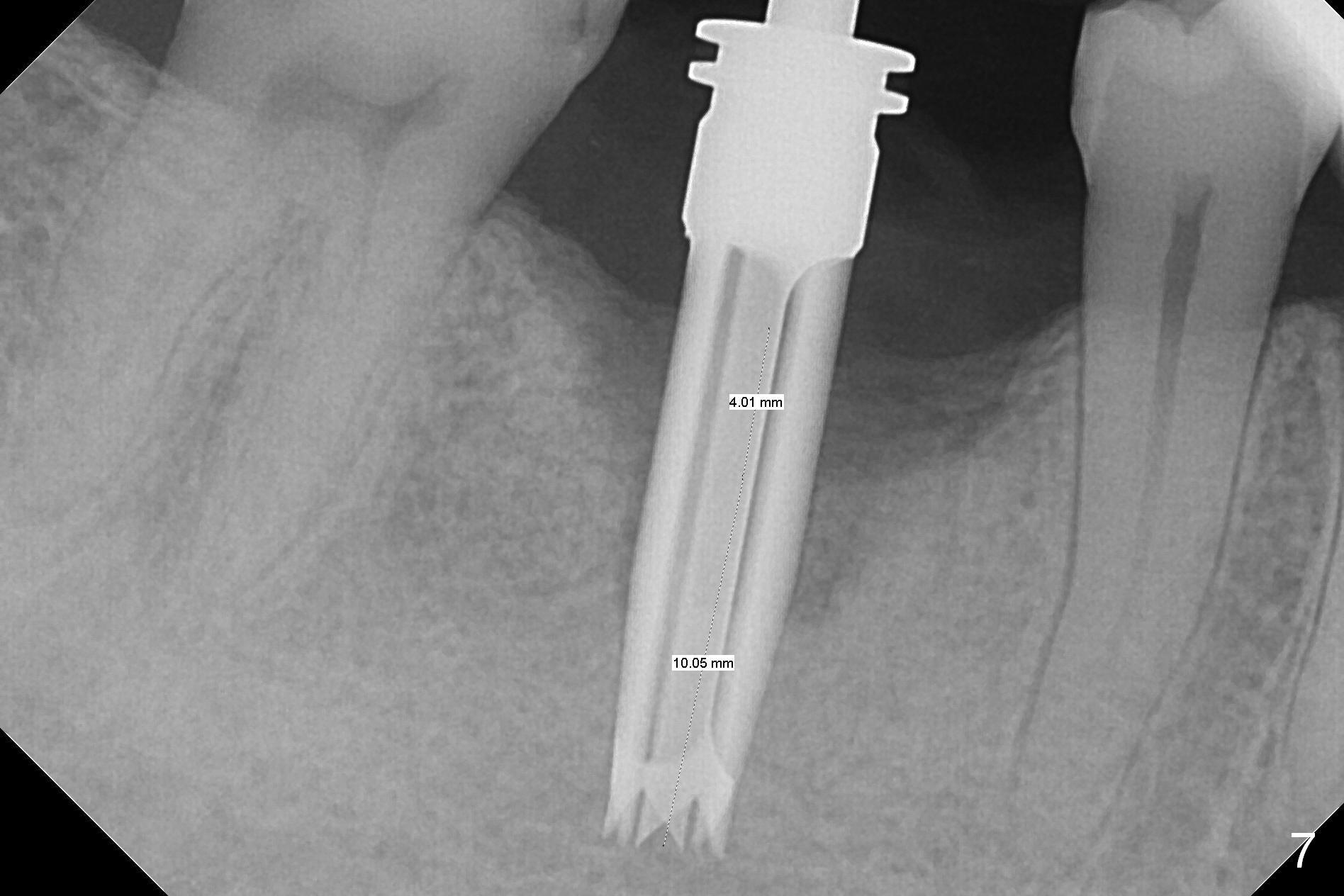
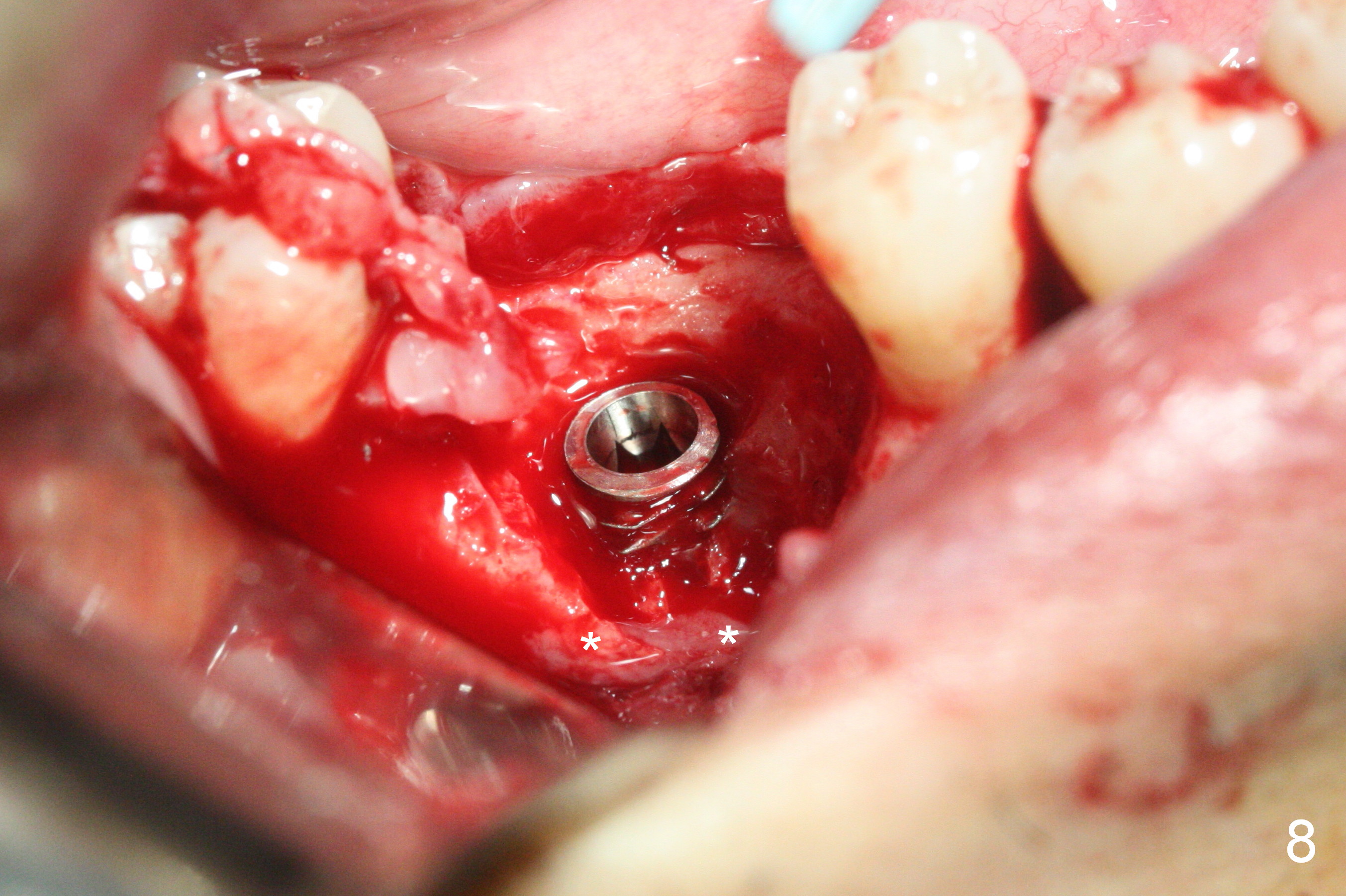
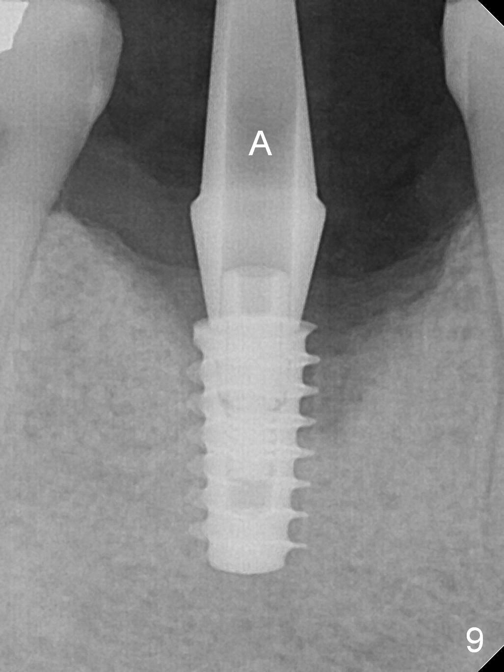
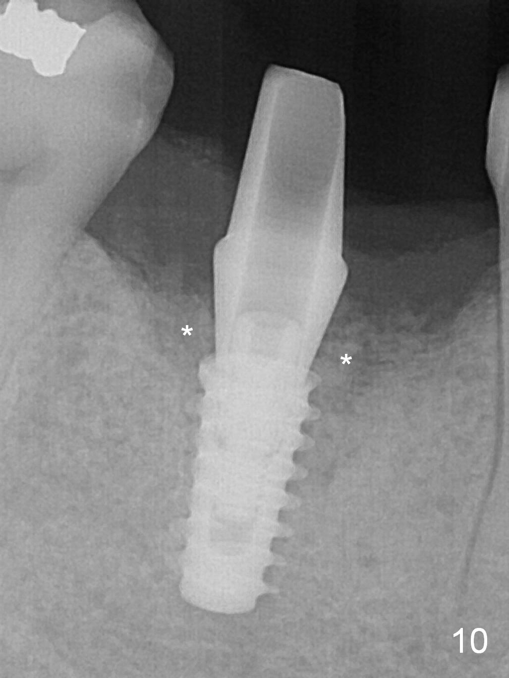
A 49-year-old man develops acute abscess of the lower right quadrant (Fig.3-5) secondary to periimplantitis at #30 (Fig.1,2,6). The latter is probably due to buccal placement (Fig.6 (*: buccal plate)). After removal of the infected implant, an osteotomy is established as lingual as possible using single drill modality (Fig.7 (4.3 mm Magic Drill after 1.6 mm pilot drill and Marking Bur, then Final Drill). With the single drill, the osteotomy does not shift buccally in spite of the lower buccal plate. A 5x9 mm IBS implant is placed lingually as planned (Fig.8 (>40 Ncm)), followed by an angled abutment (5 mm x 15° (4 mm)) (Fig.9 A). The buccal gap is filled with autogenous bone, allograft (.5-1.5 mm) and Osteogen (Fig.10 *), covered by resorbable and non-resorbable membranes. After suturing, periodontal dressing is applied.
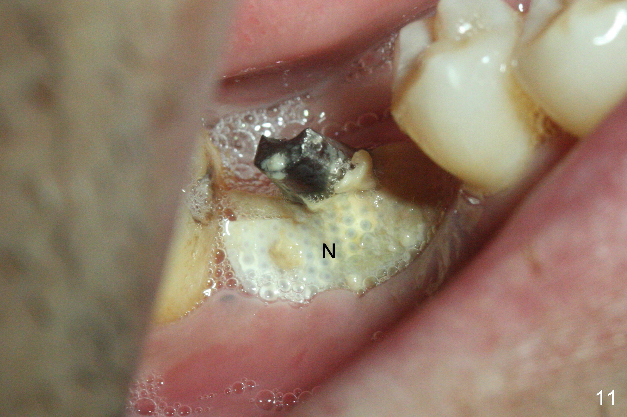
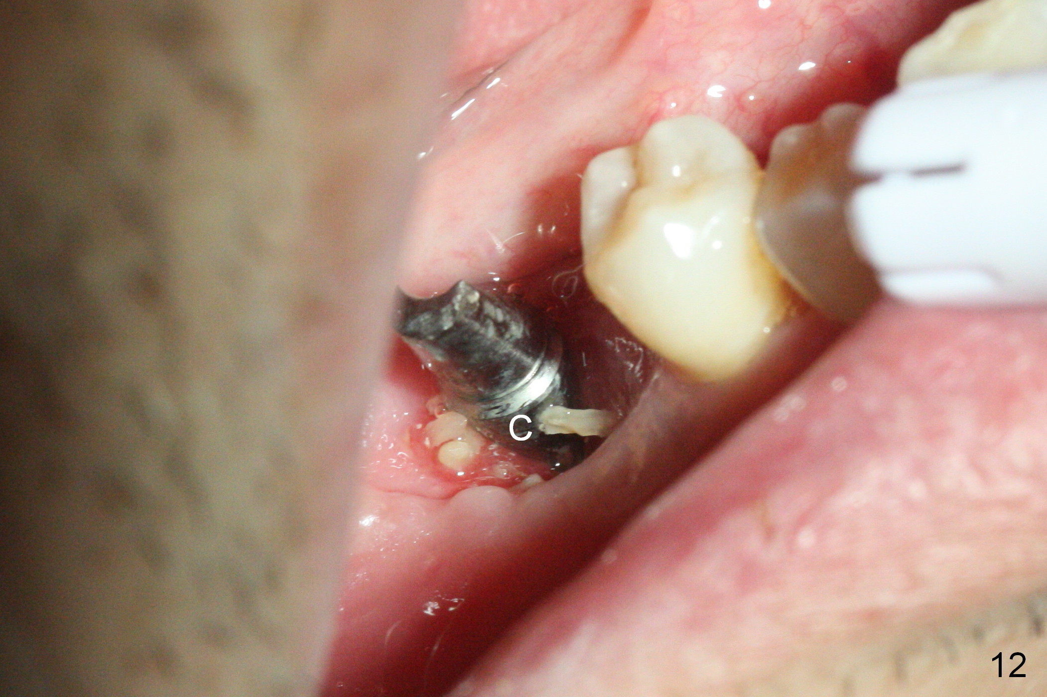
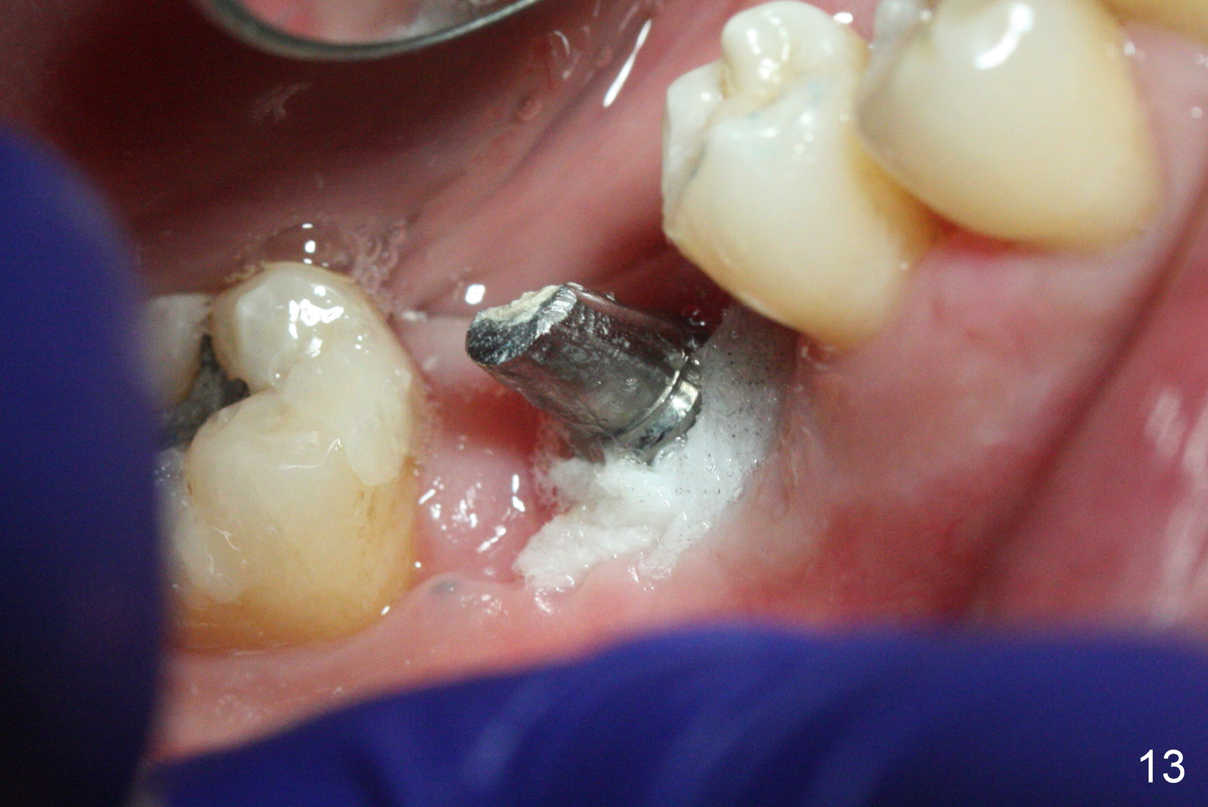
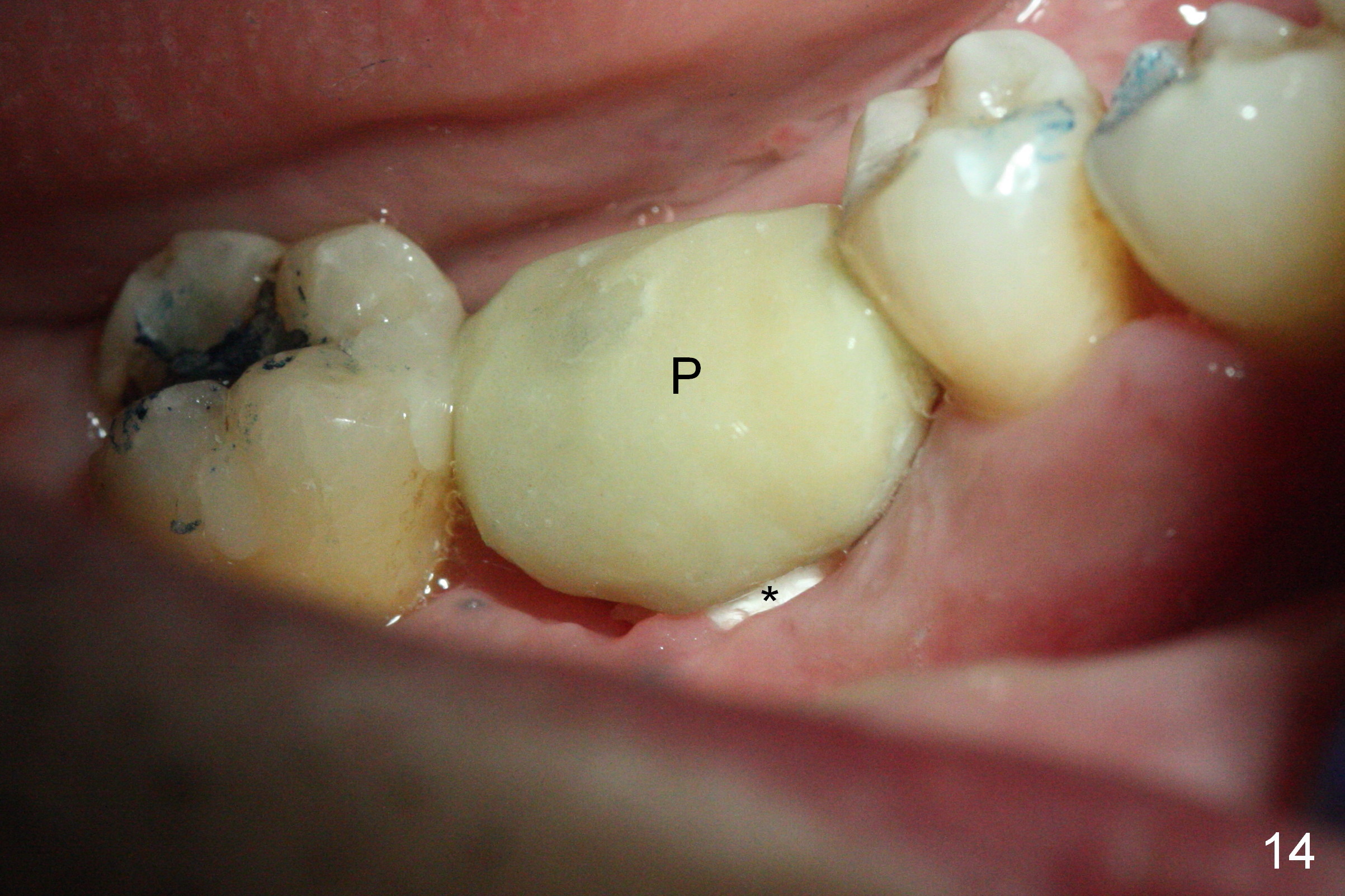
The non-resorbable membrane stays in place 1 month postop (Fig.11 N). When it is removed, the buccal aspect of the cuff of the angled abutment is not covered by bone graft or granulation tissue (Fig.12 C). In fact, the membrane does not hold the bone graft in place as effectively as a provisional. The space is filled with a piece of gauze (Fig.13) while the abutment is reduced for provisional and MO composite at #31 is placed. Before seating the provisional (Fig.14 P), a piece of collagen plug is inserted into the space (*) after removal of the gauze.
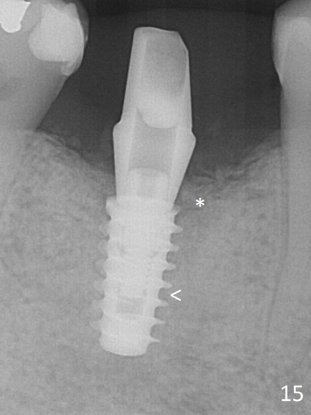
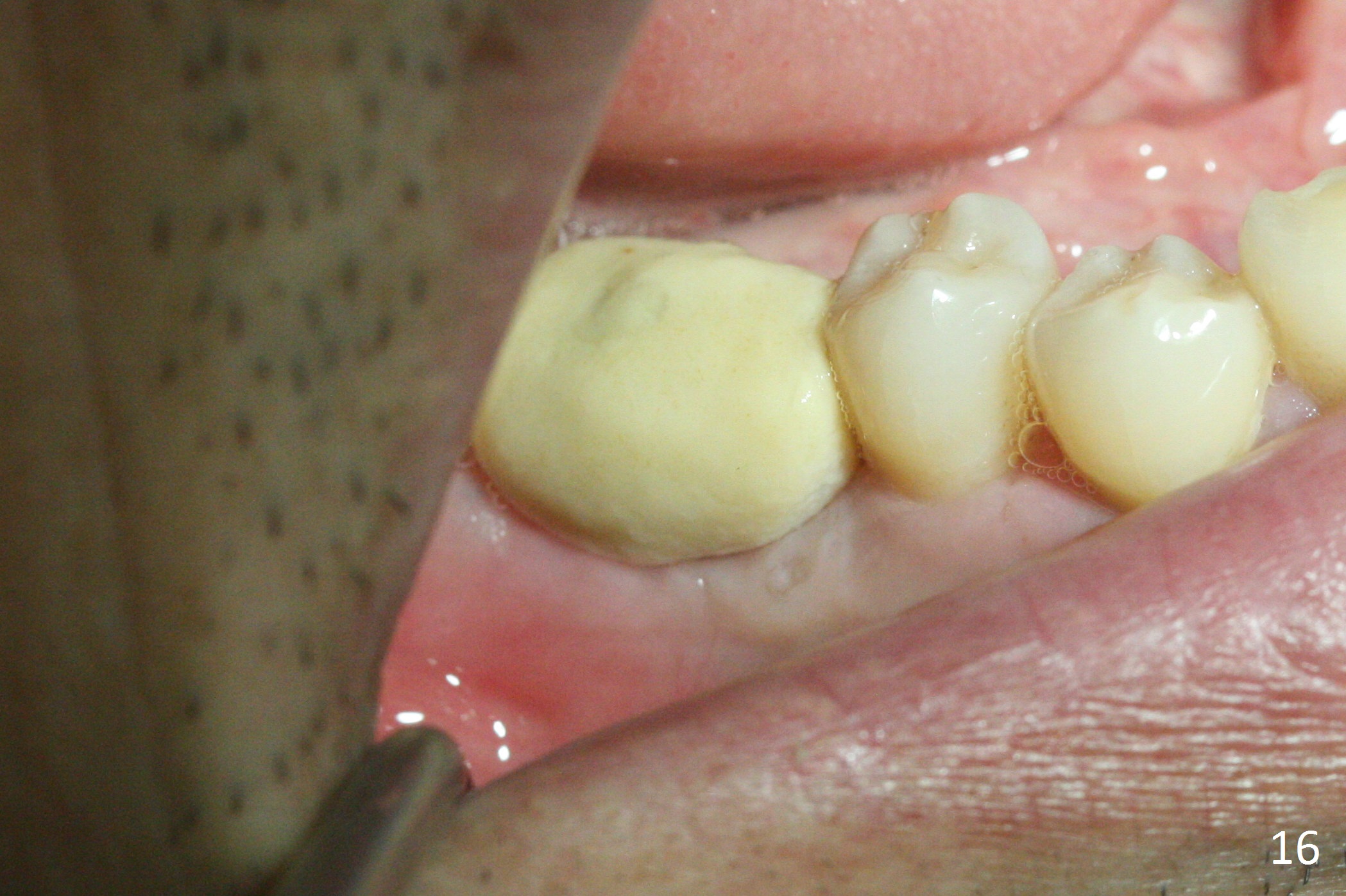
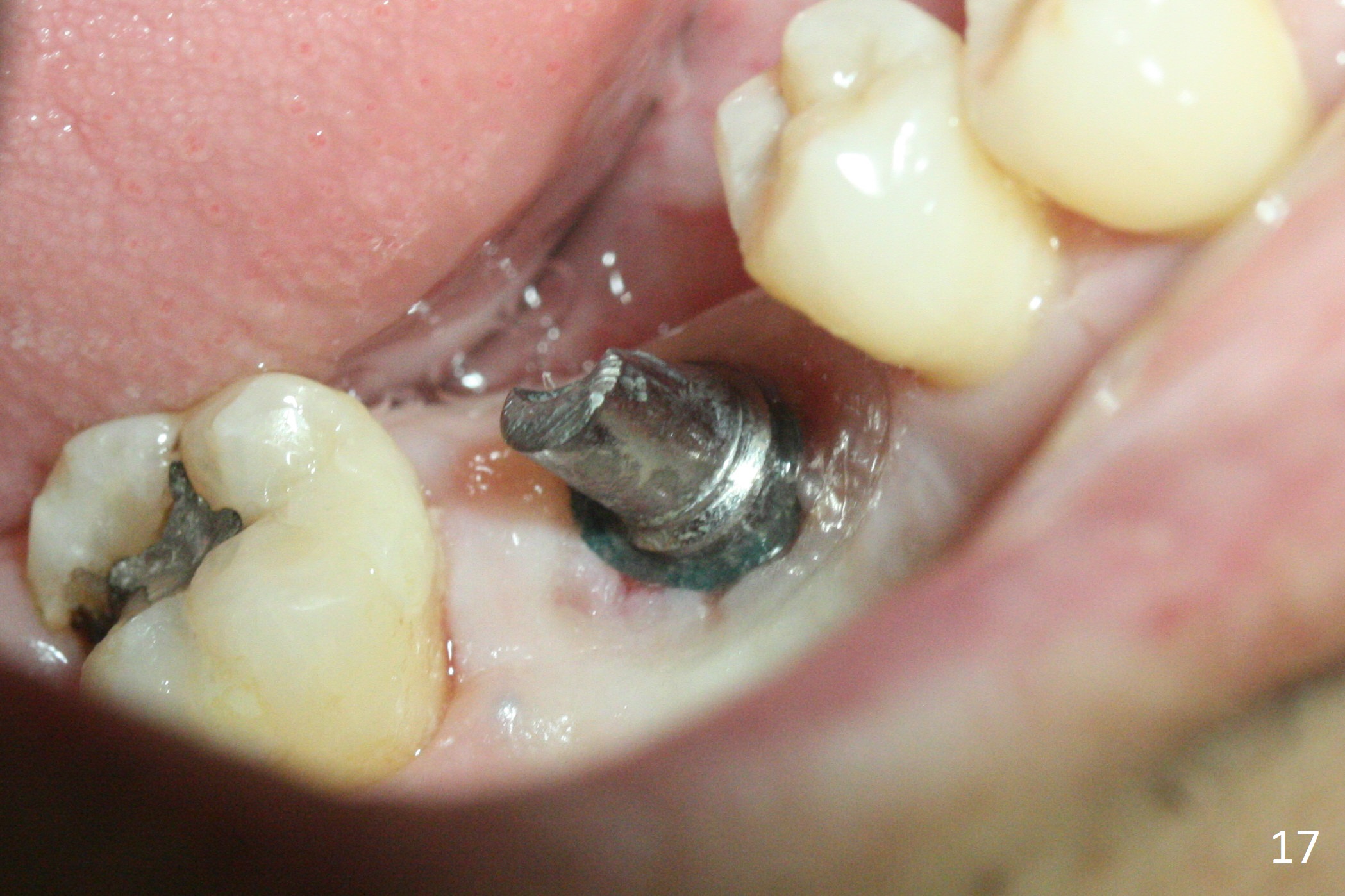
Implant threads appear to have been covered by bone coronally (Fig.15 *) and apically (<) 5 months postop. The provisional remains in place (Fig.16), although dislodged twice and replaced by the patient. The gingiva around the provisional and abutment is healthy (Fig.17 after packing with gingival retraction cord for impression).
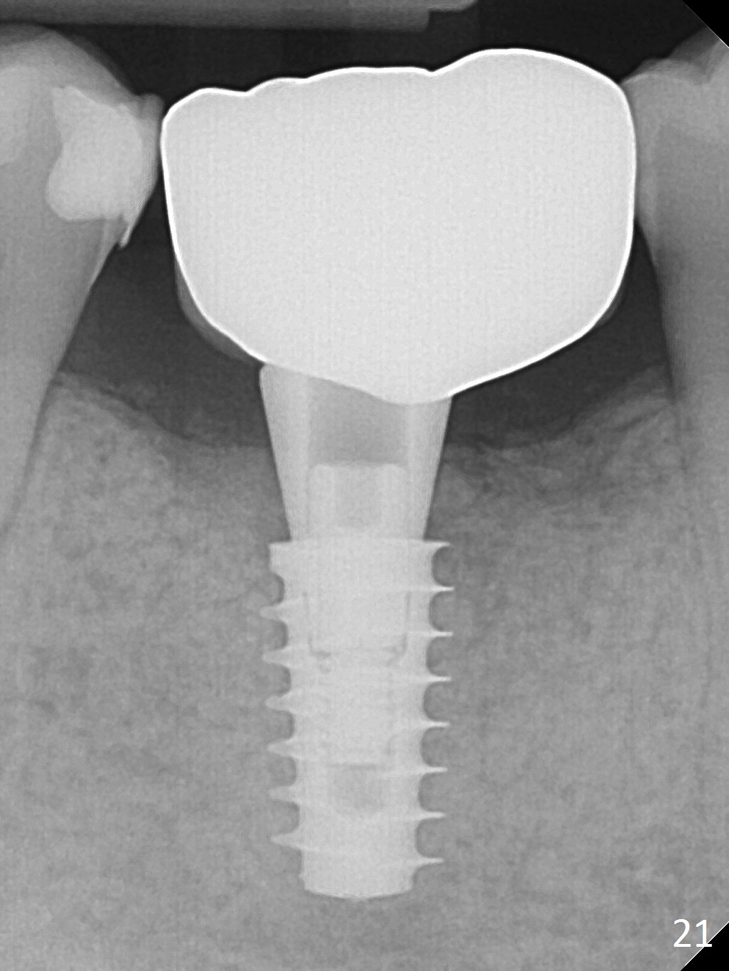
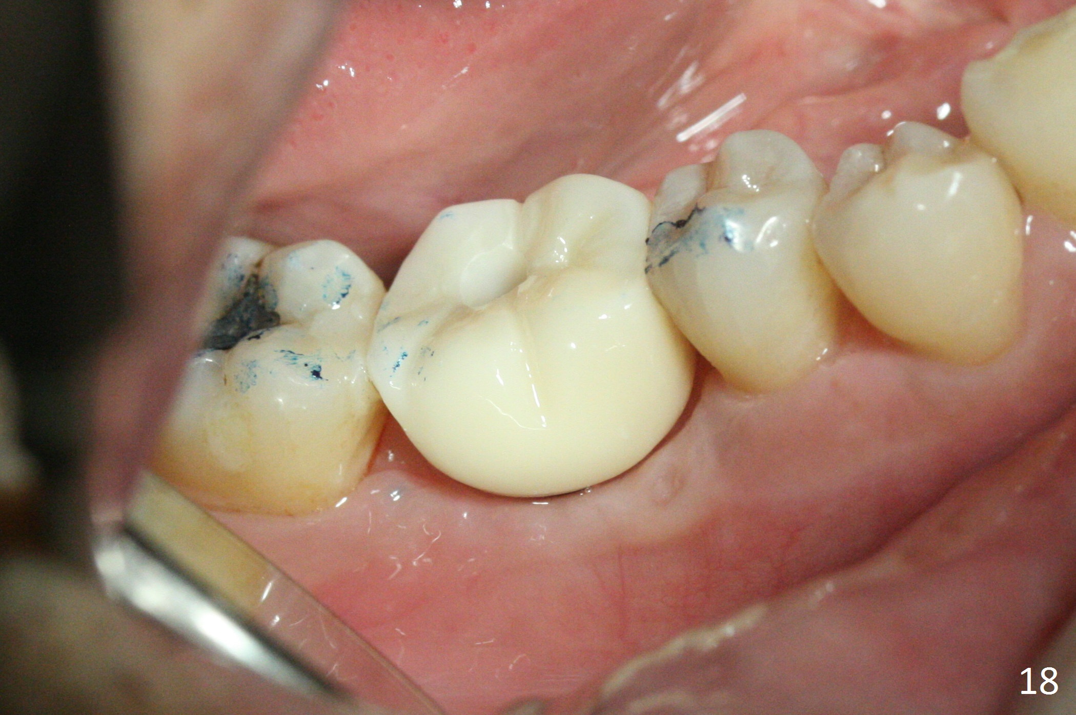
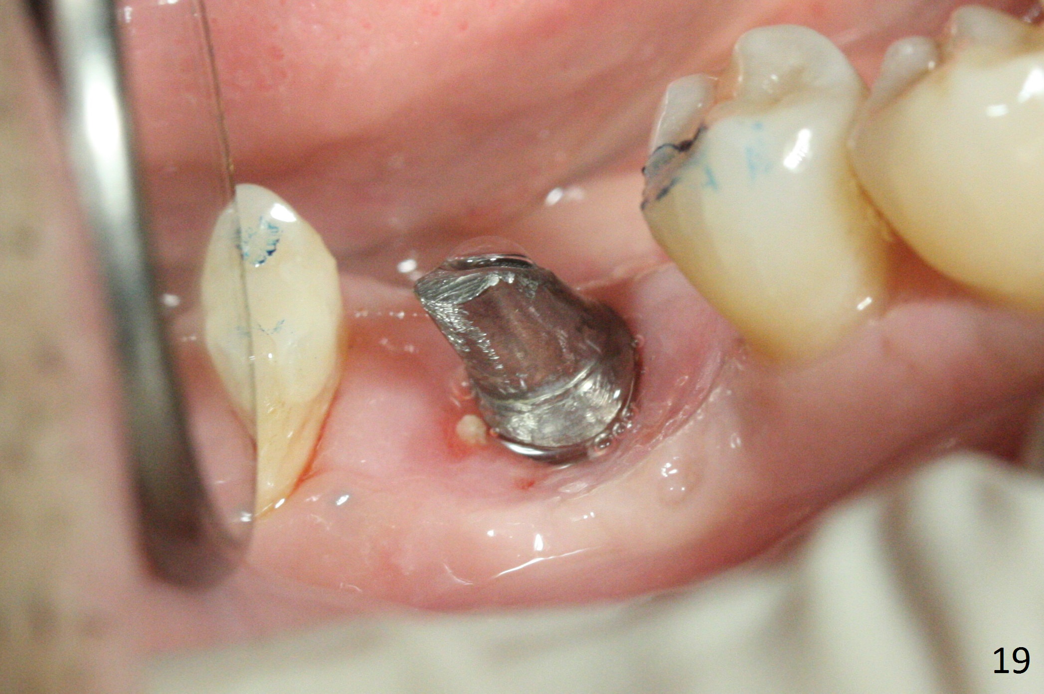
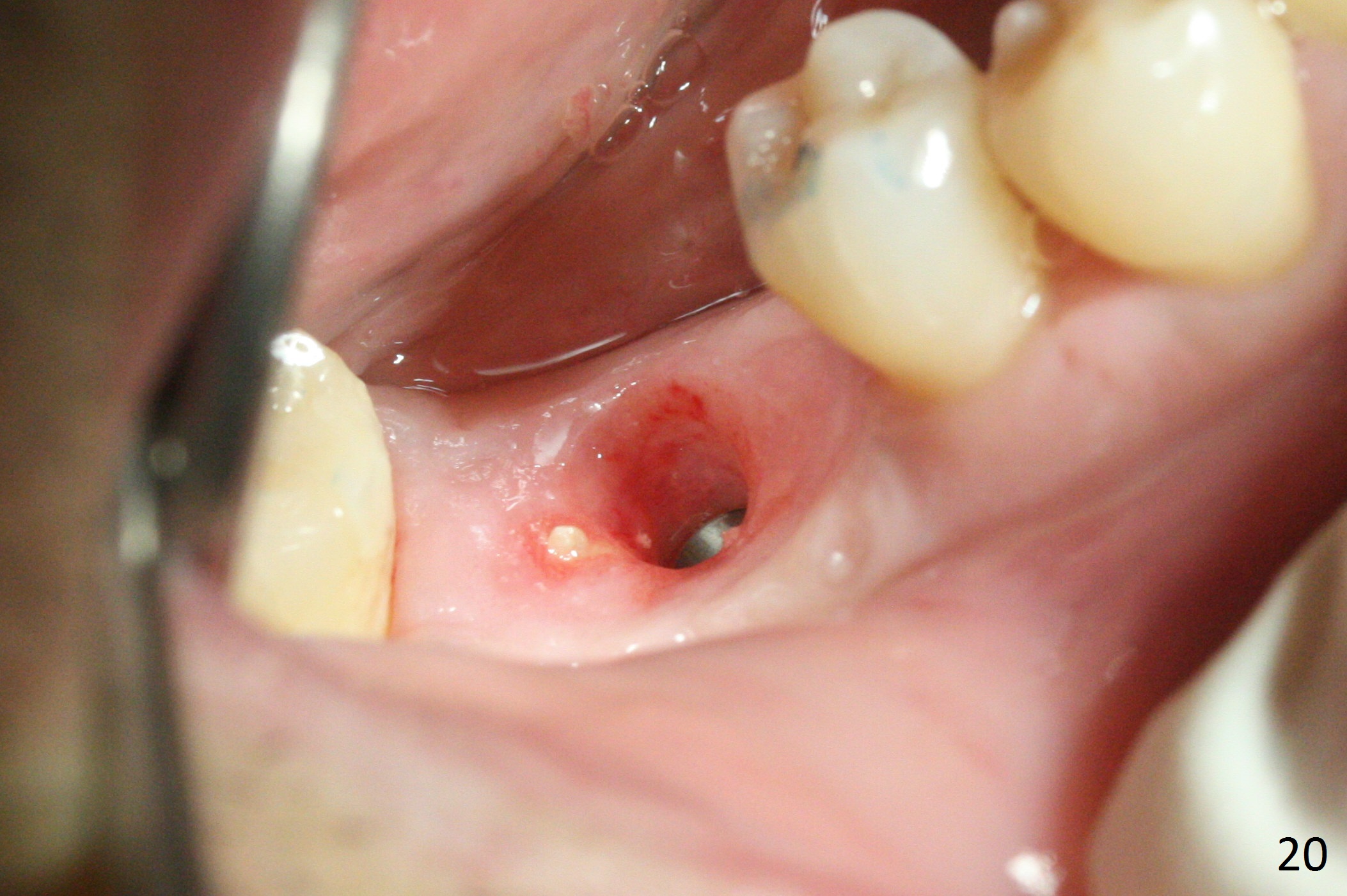
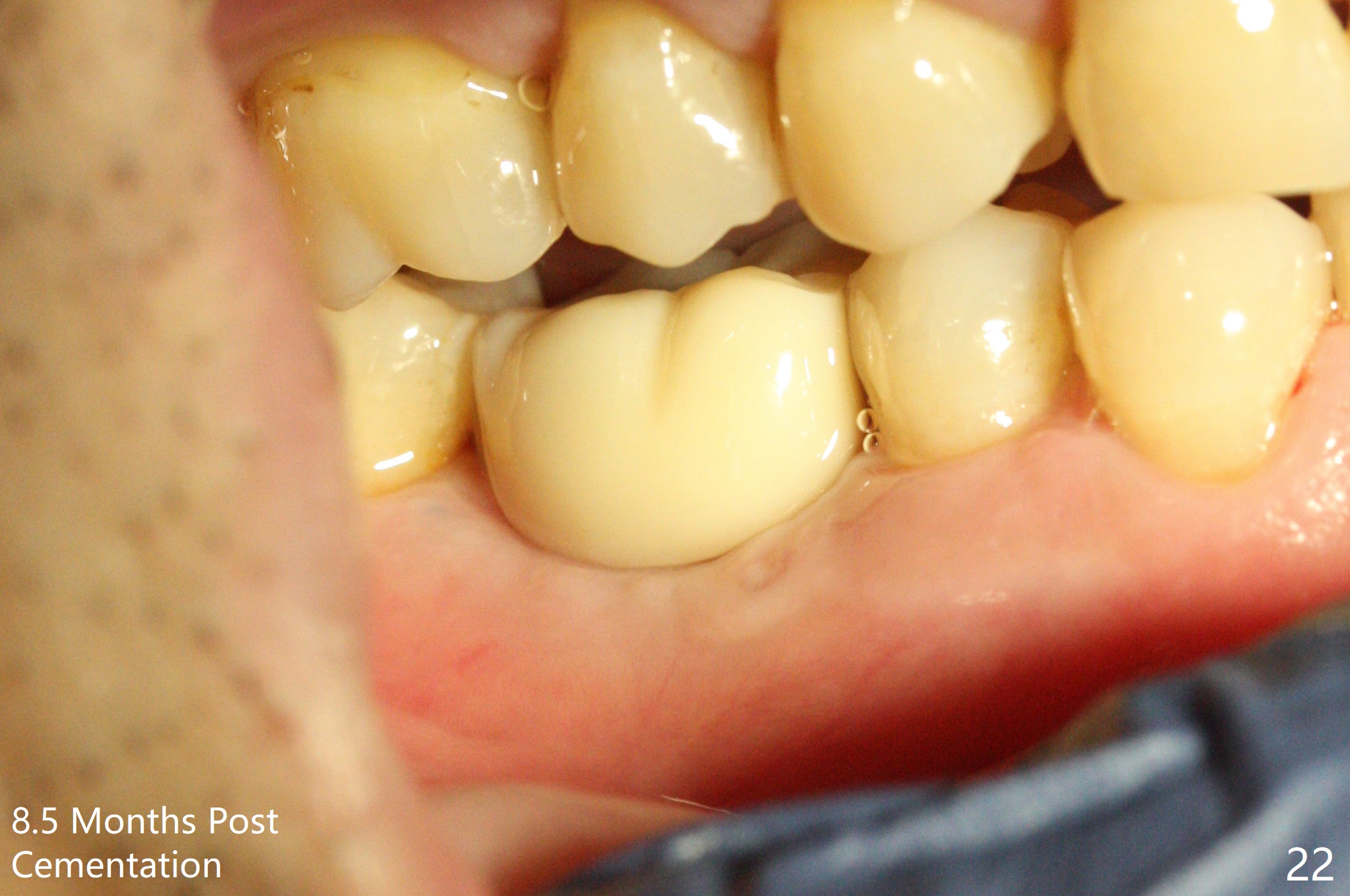
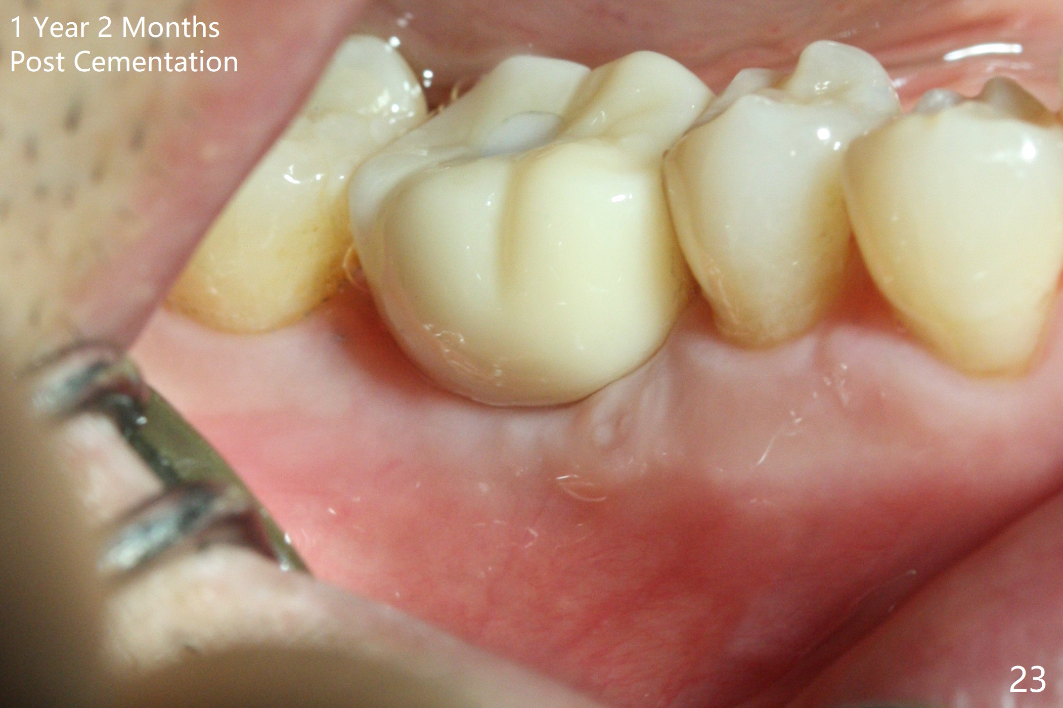
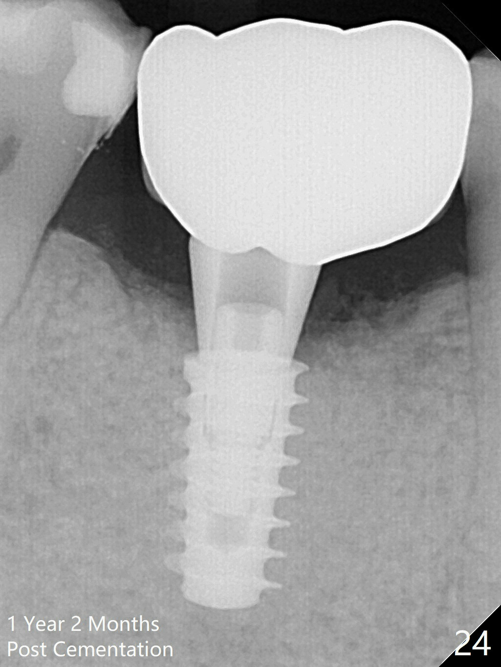
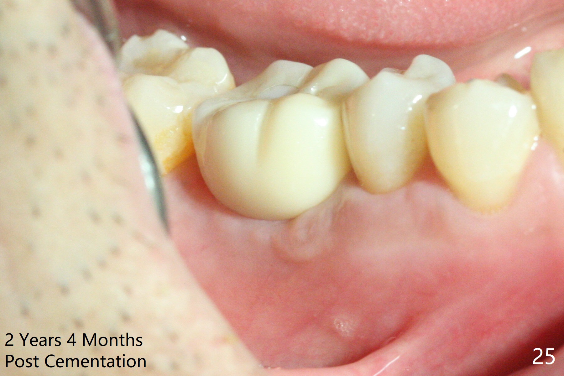
Three weeks later (5.5 months postop), a permanent crown tries in with healthy keratinized gingiva (Fig.18). The provisional keeps normal gingival bed (Fig.19), while the abutment forms tissue cuff (Fig.20). After cementation, PA shows bone regeneration (Fig.21). The gingiva remains healthy 8.5 months post cementation (Fig.22). Bone density around the implant increases 1 year 2 months post cementation (Fig.24). The gingiva remains healthy 2 years 4 months post cementation (Fig.25).
Return to
Lower
Molar Immediate Implant,
19,
Redo,
1-Year, Course
1
2
3
4
5
New Case
开场白
IBS/Guide
Xin Wei, DDS, PhD, MS 1st edition 12/19/2016, last revision 05/23/2021