
.jpg)
|
|
|
|
|
|
|
|
|
|
|
|
|
|
|
Incomplete Abutment Seating
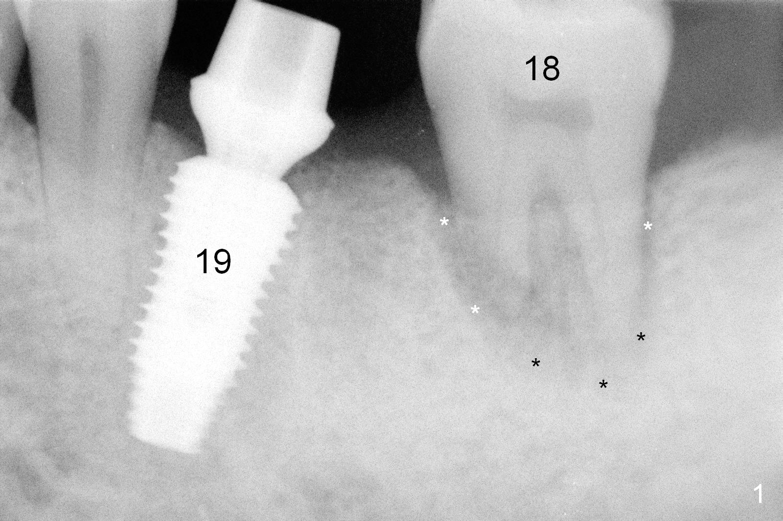
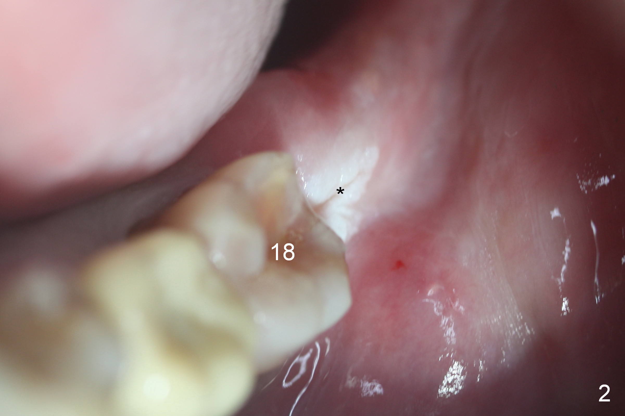
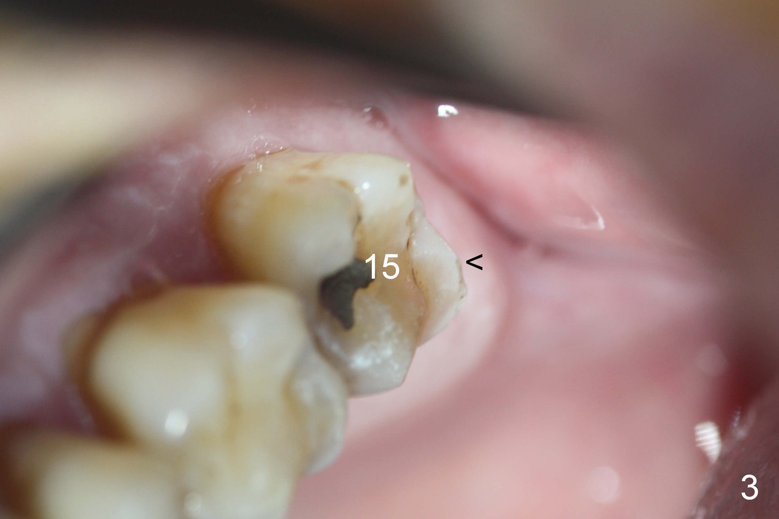
The 51-year-old patient is a severe bruxer, manifested by root resorption of the tooth #18 (Fig.1 (PA taken 8 months earlier)) with increased periodontal ligament (*: in fact fibrotic granulation tissue, apparently as a cushion). The 2nd manifestation of bruxism is the retromolar leukoplakia (Fig.2 *), corresponding to the sharp distal marginal ridge of the tooth #15 (Fig.3 <). The latter is smoothened immediately preoperatively.
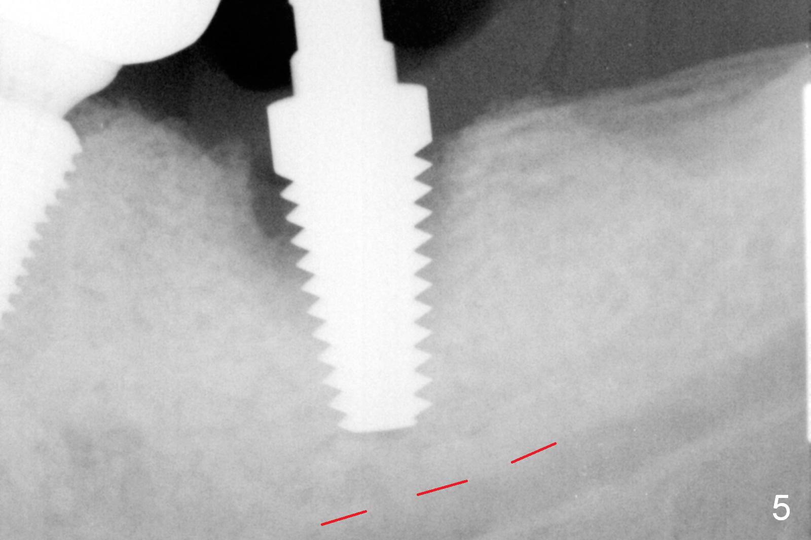
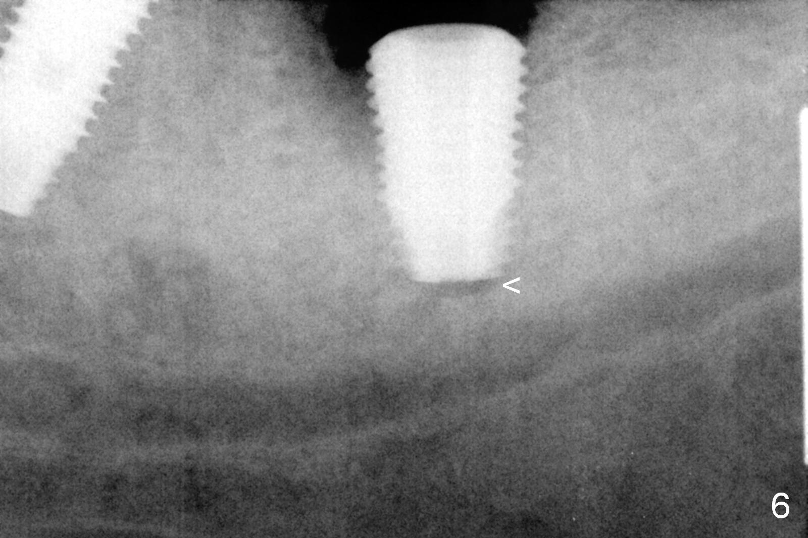
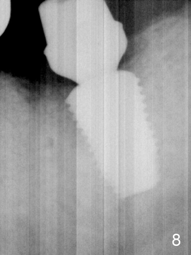
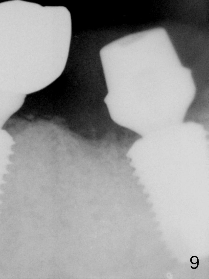
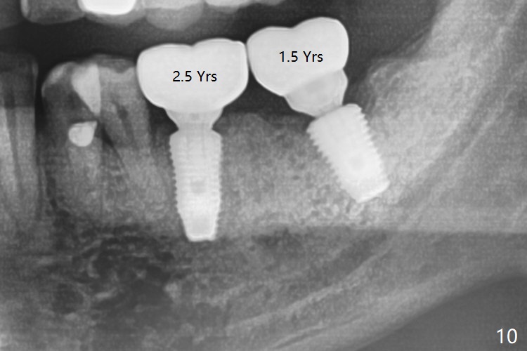
After extraction, removing the fibrotic granulation tissue fairly evenly distributed in the socket is painstaking. There is no septum left. The initial osteotomy is 3 mm apical to the bottom of the socket (Fig.4 arrow (red dashed line: the superior border of the Inferior Alveolar Canal)). The osteotomy is increased by using reamers until 4.5 mm in diameter, followed by a 7x17 mm tap (Fig.5). The osteotomy is further enlarged with 6 and 6.5 mm extra wide drills as well as 6.5 mm tap before placement of a 7x10 mm bone-level implant (Fig.6, ~ 2 mm apical to the crest). The implant is then placed deeper. A 7.5x4(4) mm cemented abutment is tried in with sufficient occlusal clearance for an immediate provisional. The abutment is removed and a small piece of gauze is inserted into the implant well prior to placement of bone graft (Fig.7 *). When the abutment is reseated, PA is taken. After placing cotton pellet and Cavit in the abutment, the occlusal clearance is insufficient. It is due to incomplete seating of the abutment (Fig.7 A). After reseating, the clearance regains and an immediate provisional is fabricated without centric and lateral occlusal contact. The bone density near the mesial and distal crests seems to increase 3.5 months postop (Fig.8,9). There appears no bone loss at the sites of #18 and 19 (1.5 and 2.5 years post cementation, respectively, Fig.10). The crown/abutment at #18is loose 4 years 8 months post cementation. It is difficult to determine whether the crown/abutment is seated complete or not because of the thick implant. It is true with a small abutment. After a week of a healing abutment, the crown/abutment is torqued at 30 Ncm smoothly, suggesting possible complete seating. Pickup impression is taken after occlusal adjustment/reduction at #18.
Return to
Lower Molar Immediate Implant
Torque
Xin Wei, DDS, PhD, MS 1st edition 04/12/2016, last revision 04/14/2021