,%206.5x4(2).jpg)
,%20hand%20tightened.jpg)
|
|
|
|
|
|
|
|
|
|
Conversion of 1st Molar to a Premolar
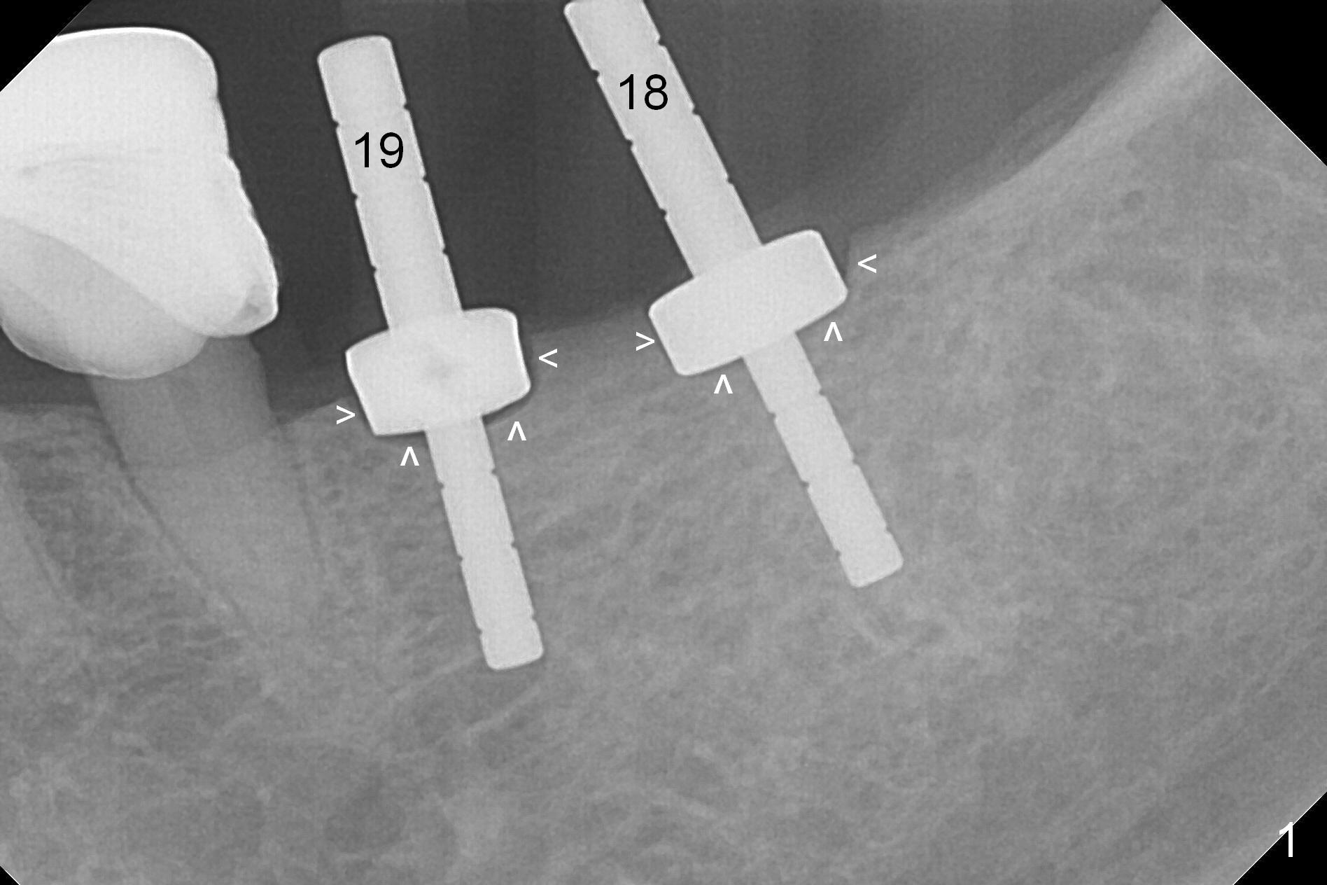
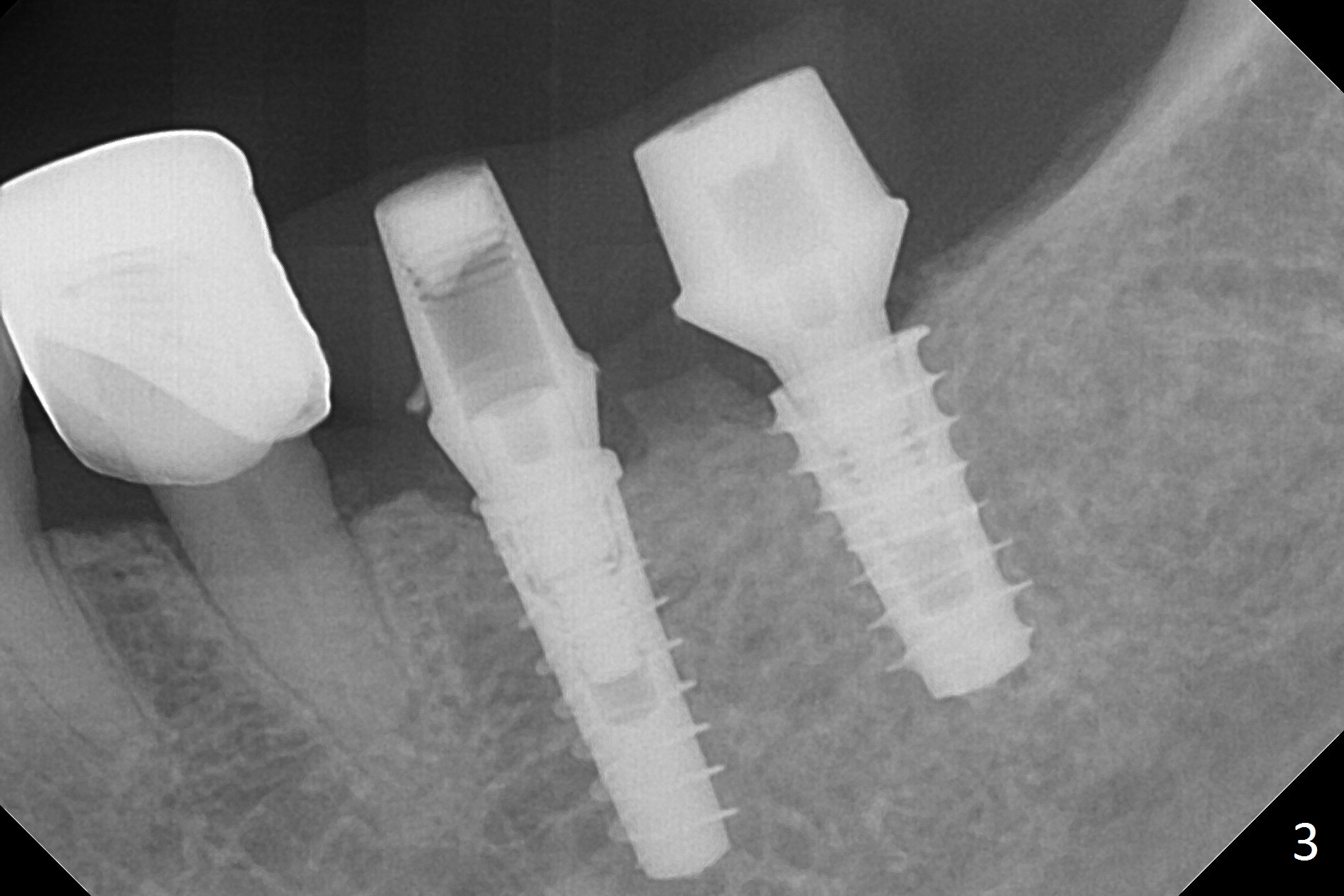
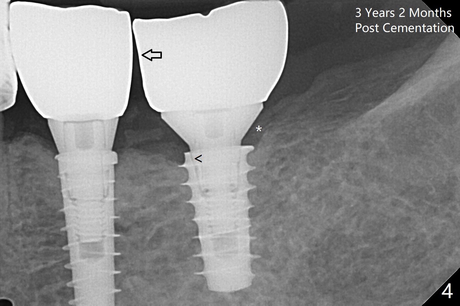
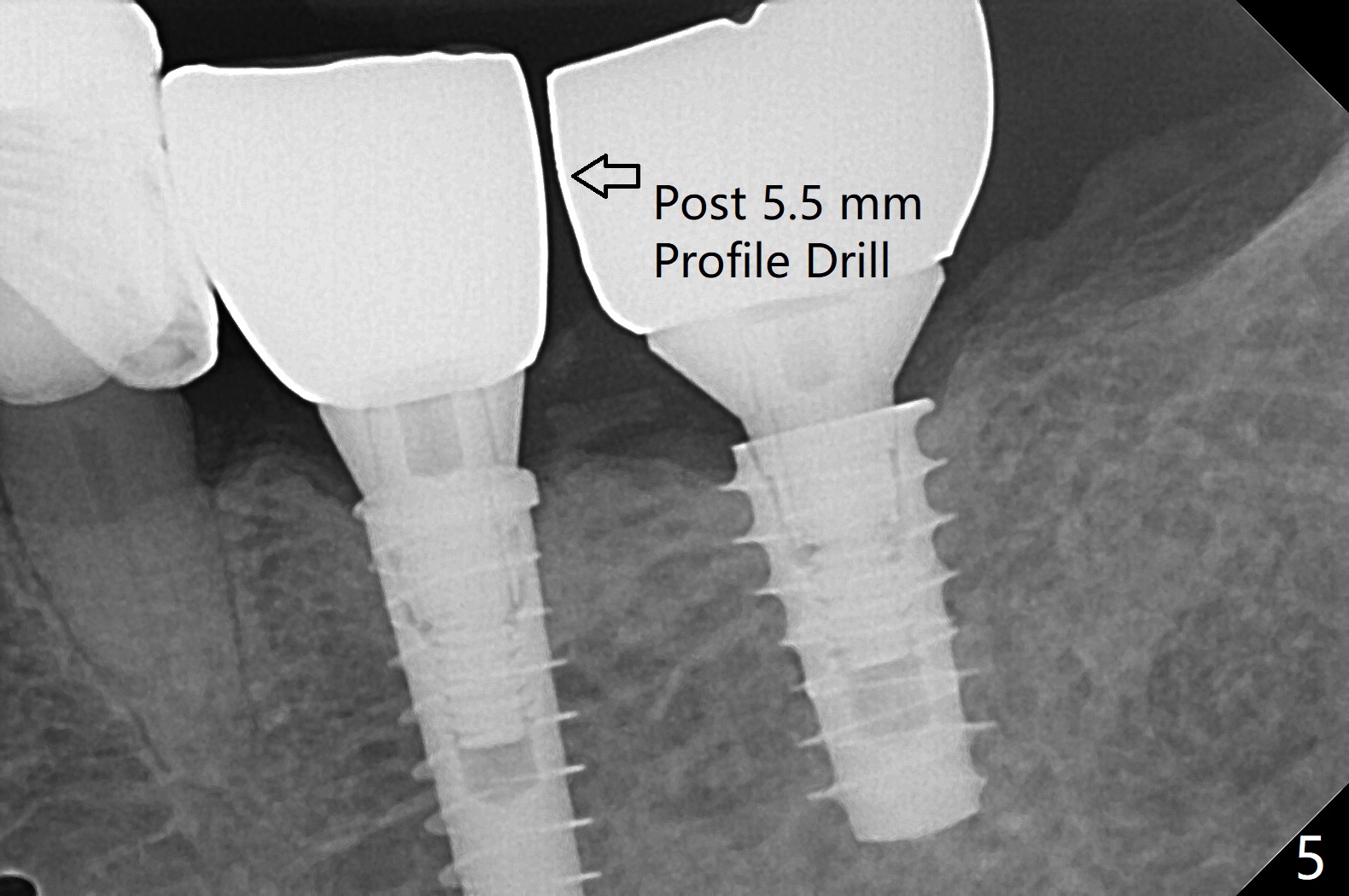
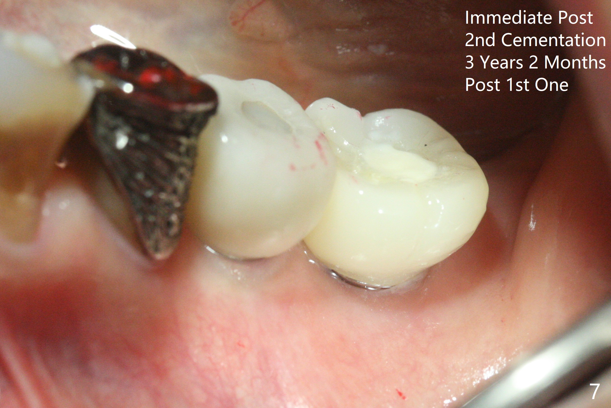
Because of the narrow ridge top at the sites of #19 and 18, it is reduced (Fig.1 arrowheads) prior to initial osteotomy with 1.6 mm pilot drill. After Magic Drills (3.3 and 4.3 mm at #19 and 18, respectively), 4x11 and 5x9 mm IBS implants are placed with insertion torque >35 Ncm with immediate placement of pair abutments (4.5x5.7(2) and 6.5x4(2) mm, Fig.2). In fact these sites are converted to a premolar and a 1st molar (because narrow ridge at #19). Following GBR and suturing, periodontal dressing is applied around the abutments for increased retention. The regional ridge reduction makes Marking Bur unnecessary (because of flat ridge top and the soft bone in this case) and more importantly there is no thread exposure upon implant placement. The trimmed site (concavity) is favorable for bone graft and membrane placement. One month postop, loose perio dressing is removed and replaced by a splinted nonfunctional provisional. The implant sites look normal nearly 3 months postop; there is no bone loss (Fig.3). Impression is taken. The crown/abutment at #18 is loose 3 years 2 months post cementation; when the crown/abutment is retightened, the abutment remains incompletely seated (Fig.4 <) in spite of reduction of the proximal contact (arrow). It may be due to the block of the distal crest (*). After use of 5.5 mm profile drill, the 6.5x4(2) mm abutment remains unseated (Fig.5). The smaller one (5x4(2) mm, Fig.6) is seated. When the redo crown is cemented, the surrounding gingiva is healthy with a short papilla between the implant crowns (Fig.7).
Lower Molar Immediate Implant 29-31 CT Post Cementation Coronal Section
Xin Wei, DDS, PhD, MS 1st edition 01/26/2017, last revision 10/01/2020