|
|
|
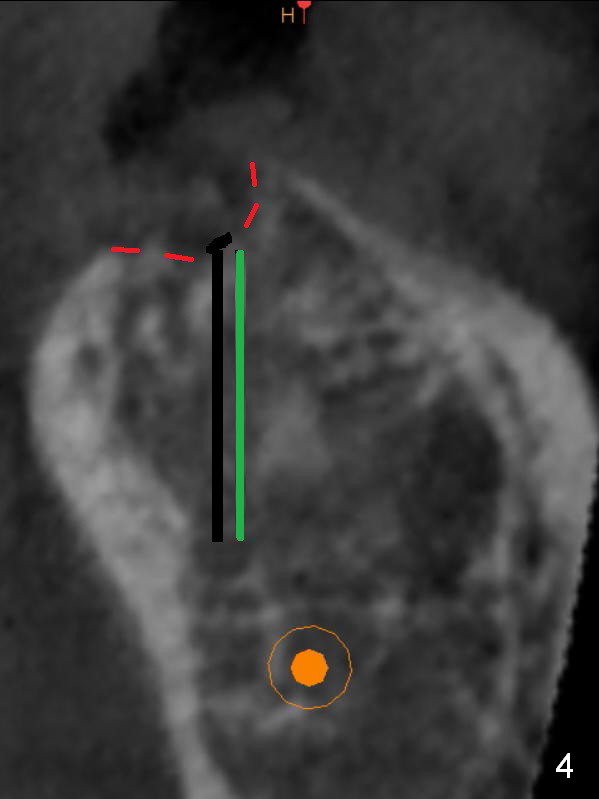
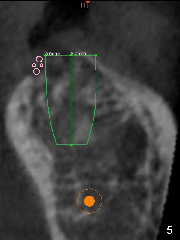
Osteotomy is initiated in the middle of the concavity (Fig.2 green line) with insertion of 7 mm guide pin (Fig.3). After removal of the pin, the osteotomy is moved buccally (Fig.4). After Marking Bur and 4.3 mm Magic Drill, a 5x9 mm IBS implant is placed with 2.8 mm clearance from the Inferior Alveolar Canal (Fig.6). Following deepening the osteotomy with Final Drill, the implant is placed deeper (Fig.7). The osteotomy happens to be established in the mesial socket, since the distal socket has not completely healed (Fig.6 yellow dashed line). Granulation tissue is removed. Since the lingual crest is lower than the buccal one (Fig.1 B), there is lingual thread exposure after implant placement (Fig.5). The exposed thread is covered by bone graft (autogenous bone, allograft and Osteogen, Fig.5 pink circles). Mesial Socket Placement Last Next Xin Wei, DDS, PhD, MS 1st edition 01/20/2017, last revision 08/30/2018